Japanese
English
- 有料閲覧
- Abstract 文献概要
- 1ページ目 Look Inside
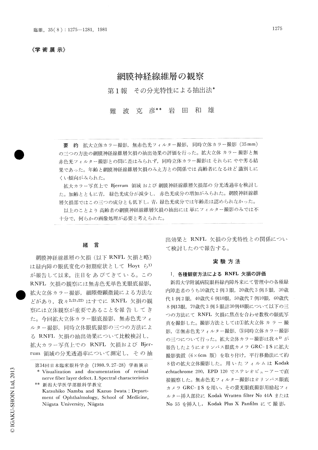
拡大立体カラー撮影,無赤色光フィルター撮影,同時立体カラー撮影(35mm)の三つの方法の網膜神経線維層欠損の抽出効果の評価を行った。拡大立体カラー撮影と無赤色光フィルター撮影との間に差はみられず,同時立体カラー撮影はそれらにやや劣る結果であった。年齢と網膜神経線維層欠損のみえ方との関係では高齢者になるほど識別しにくい傾向がみられた。
拡大カラー写真上でBjerrum領域および網膜神経線維層欠損部の分光透過率を検討した。加齢とともに青,緑色光成分が減少し,赤色光成分の増加がみられた。網膜神経線維層欠損部ではこの三つの成分とも低下し,青,緑色光成分では年齢差は認められなかった。
以上のことより高齢者の網膜神経線維層欠損の抽出には単にフィルター撮影のみでは不十分で,何らかの画像処理が必要と考えられた。
We attemped to evaluate the photographic methods for the retinal nerve fiber layer defect (RNFL-D) in glaucomatous eyes, comparing the fundus photographs obtained by the following three methods: magnified stereo-color fundus pho-tography, monochromatic fundus photography using Kodak Wratten filter No 44 A or No 55 and simultaneous stereo-color fundus photography. The result showed no significant difference be-tween the magnified stereo-color fundus photo-graphy and monochromatic photography and both had advantages over the simultaeneous stereophotography.

Copyright © 1981, Igaku-Shoin Ltd. All rights reserved.


