Japanese
English
- 有料閲覧
- Abstract 文献概要
- 1ページ目 Look Inside
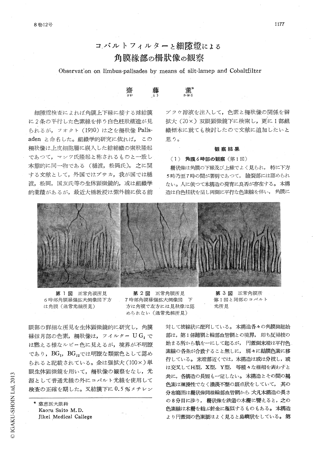
細隙燈検査によれば角膜上下縁に接する球結膜に2条の平行した色素線を伴う白色柱状構造が見られるが,フオクト(1930)は之を柵状像Palis-adenと命名した。組織学的研究に依れば,この柵状像は上皮細胞層に嵌入した結締織の索状隆起であつて,マンツ氏隆起と称されるものと一致し本態的に同一物である(樋渡,松岡氏)。之に関する文献として,外国ではブサカ,我が国では樋渡,松岡,国友氏等の生体顕微鏡的,或は組織学的業蹟があるが,最近大橋教授は紫外線に依る前眼部の詳細な所見を生体顕微鏡的に研究し,角膜縁弦月部の色素,柵状像は,フイルターUG1では燃える様なルビー色に見えるが,境界が不明瞭であり,BG1,BG12では明瞭な類紫色として認められると記載されている。余は強拡大(100×)単眼生体顕微鏡を用いて,柵状像の観察をなし,光源として普通光線の外にコバルト光線を使用して検査の正確を期した。又結膜下に0.5%メチレンブラウ溶液を注入して,色素と柵状像の関係を弱拡大(20×)双眼顕微鏡下に検索し,更に1部組織標本に就ても検討したので文献に追加したいと思う。
Observations of limbus-palisades by means of bio-microscope aided by slit-lamp and cobalt-filter are quite in agreement with what Vogt has already described. For this purpose Cob-alt-filter is an important adjunct; the use of bio-dying brings out limbus-palisades vividly to view. The probable function of the structure in question, the author belives, is an agent whereby, the arterioles and venules that lie in the limbus which serves as the passage-way for the lymph of the eyeball as well as for the fluids that provide corneal nutrition are given protection.

Copyright © 1954, Igaku-Shoin Ltd. All rights reserved.


