Japanese
English
- 有料閲覧
- Abstract 文献概要
- 1ページ目 Look Inside
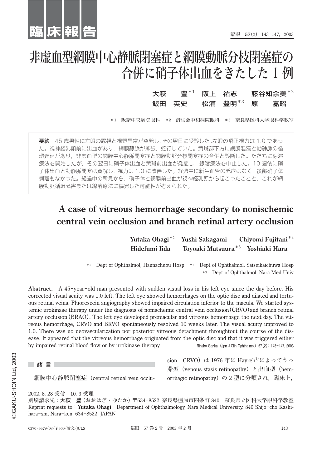
要約 45歳男性に左眼の霧視と視野異常が突発し,その翌日に受診した。左眼の矯正視力は1.0であった。視神経乳頭前に出血があり,網膜静脈が拡張,蛇行していた。黄斑部下方に網膜混濁と動静脈の循環遅延があり,非虚血型の網膜中心静脈閉塞症と網膜動脈分枝閉塞症の合併と診断した。ただちに線溶療法を開始したが,その翌日に硝子体出血と黄斑前出血が発症し,線溶療法を中止した。10週後に硝子体出血と動静脈閉塞は寛解し,視力は1.0に改善した。経過中に新生血管の発症はなく,後部硝子体剝離もなかった。経過中の所見から,硝子体と網膜前出血が視神経乳頭から起こったことと,これが網膜動脈循環障害または線溶療法に続発した可能性が考えられた。
Abstract. A 45-year-old man presented with sudden visual loss in his left eye since the day before. His corrected visual acuity was 1.0 left. The left eye showed hemorrhages on the optic disc and dilated and tortuous retinal veins. Fluorescein angiography showed impaired circulation inferior to the macula. We started systemic urokinase therapy under the diagnosis of nonischemic central vein occlusion(CRVO)and branch retinal artery occlusion(BRAO). The left eye developed premacular and vitreous hemorrhage the next day. The vitreous hemorrhage,CRVO and BRVO spontaneously resolved 10 weeks later. The visual acuity improved to 1.0. There was no neovascularization nor posterior vitreous detachment throughtout the course of the disease. It appeared that the vitreous hemorrhage originated from the optic disc and that it was triggered either by impaired retinal blood flow or by urokinase therapy.

Copyright © 2003, Igaku-Shoin Ltd. All rights reserved.


