Japanese
English
- 有料閲覧
- Abstract 文献概要
- 1ページ目 Look Inside
- 参考文献 Reference
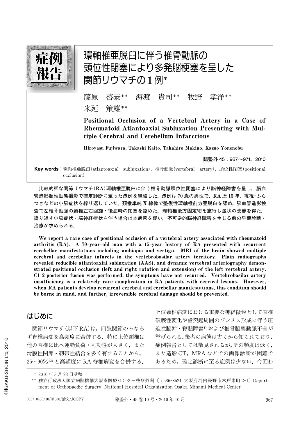
比較的稀な関節リウマチ(RA)環軸椎亜脱臼に伴う椎骨動脈頭位性閉塞により脳神経障害を呈し,脳血管造影頚椎動態撮影で確定診断に至った症例を経験した.症例は70歳の男性で,RA歴15年,複視・ふらつきなどの小脳症状を繰り返していた.頚椎単純X線像で整復性環軸椎前方亜脱臼を認め,脳血管造影検査で左椎骨動脈の頚椎左右回旋・後屈時の閉塞を認めた.環軸椎後方固定術を施行し症状の改善を得た.繰り返す小脳症状・脳神経症状を伴う場合は本病態を疑い,不可逆的脳神経障害を生じる前の早期診断・治療が求められる.
We report a rare case of positional occlusion of a vertebral artery associated with rheumatoid arthritis (RA). A 70-year-old man with a 15-year history of RA presented with recurrent cerebellar manifestations including ambiopia and vertigo. MRI of the brain showed multiple cerebral and cerebellar infarcts in the vertebrobasilar artery territory. Plain radiographs revealed reducible atlantoaxial subluxation (AAS), and dynamic vertebral arteriography demonstrated positional occlusion (left and right rotation and extension) of the left vertebral artery. C1-2 posterior fusion was performed, the symptoms have not recurred. Vertebrobasilar artery insufficiency is a relatively rare complication in RA patients with cervical lesions. However, when RA patients develop recurrent cerebral and cerebellar manifestations, this condition should be borne in mind, and further, irreversible cerebral damage should be prevented.

Copyright © 2010, Igaku-Shoin Ltd. All rights reserved.


