Japanese
English
- 有料閲覧
- Abstract 文献概要
- 1ページ目 Look Inside
- 参考文献 Reference
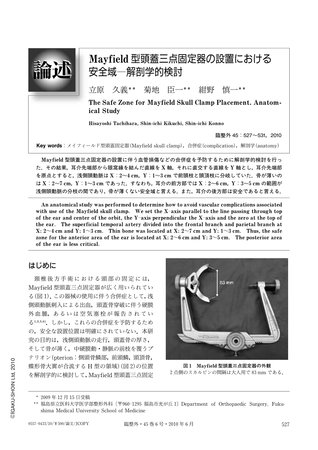
Mayfield型頭蓋三点固定器の設置に伴う血管損傷などの合併症を予防するために解剖学的検討を行った.その結果,耳介先端部から眼窩縁を結んだ直線をX軸,それに直交する直線をY軸とし,耳介先端部を原点とすると,浅側頭動脈はX:2~4cm,Y:1~3cmで前頭枝と頭頂枝に分岐していた.骨が薄いのはX:2~7cm,Y:1~3cmであった.すなわち,耳介の前方部ではX:2~6cm,Y:3~5cmの範囲が浅側頭動脈の分枝の間であり,骨が薄くない安全域と言える.また,耳介の後方部は安全であると言える.
An anatomical study was performed to determine how to avoid vascular complications associated with use of the Mayfield skull clamp. We set the X-axis parallel to the line passing through top of the ear and center of the orbit, the Y-axis perpendicular the X-axis and the zero at the top of the ear. The superficial temporal artery divided into the frontal branch and parietal branch at X: 2~4cm and Y: 1~3cm. Thin bone was located at X: 2~7cm and Y: 1~3cm. Thus, the safe zone for the anterior area of the ear is located at X: 2~6cm and Y: 3~5cm. The posterior area of the ear is less critical.

Copyright © 2010, Igaku-Shoin Ltd. All rights reserved.


