Japanese
English
- 有料閲覧
- Abstract 文献概要
- 1ページ目 Look Inside
- 参考文献 Reference
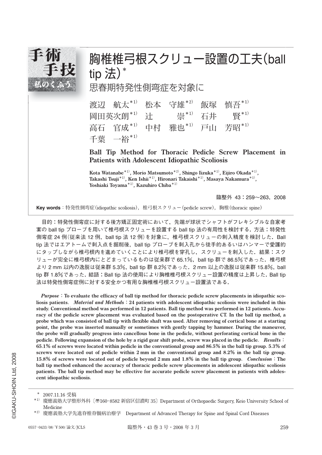
目的:特発性側弯症に対する後方矯正固定術において,先端が球状でシャフトがフレキシブルな自家考案のball tipプローブを用いて椎弓根スクリューを設置するball tip法の有用性を検討する.方法:特発性側弯症24例(従来法12例,ball tip法12例)を対象に,椎弓根スクリューの刺入精度を検討した.Ball tip法ではエアトームで刺入点を掘削後,ball tipプローブを刺入孔から徒手的あるいはハンマーで愛護的にタップしながら椎弓根内を進めていくことにより椎弓根を穿孔し,スクリューを刺入した.結果:スクリューが完全に椎弓根内にとどまっているものは従来群で65.1%,ball tip群で86.5%であった.椎弓根より2mm以内の逸脱は従来群5.3%,ball tip群8.2%であった.2mm以上の逸脱は従来群15.8%,ball tip群1.8%であった.結語:Ball tip法の使用により胸椎椎弓根スクリュー設置の精度は上昇した.Ball tip法は特発性側弯症例に対する安全かつ有用な胸椎椎弓根スクリュー設置法である.
Purpose:To evaluate the efficacy of ball tip method for thoracic pedicle screw placements in idiopathic scoliosis patients. Material and Methods:24 patients with adolescent idiopathic scoliosis were included in this study. Conventional method was performed in 12 patients. Ball tip method was performed in 12 patients. Accuracy of the pedicle screw placement was evaluated based on the postoperative CT. In the ball tip method, a probe which was consisted of ball tip with flexible shaft was used. After removing of cortical bone at a starting point, the probe was inserted manually or sometimes with gently tapping by hammer. During the maneuver, the probe will gradually progress into cancellous bone in the pedicle, without perforating cortical bone in the pedicle. Following expansion of the hole by a rigid gear shift probe, screw was placed in the pedicle. Results:65.1% of screws were located within pedicle in the conventional group and 86.5% in the ball tip group. 5.3% of screws were located out of pedicle within 2mm in the conventional group and 8.2% in the ball tip group. 15.8% of screws were located out of pedicle beyond 2mm and 1.8% in the ball tip group. Conclusion:The ball tip method enhanced the accuracy of thoracic pedicle screw placements in adolescent idiopathic scoliosis patients. The ball tip method may be effective for accurate pedicle screw placement in patients with adolescent idiopathic scoliosis.

Copyright © 2008, Igaku-Shoin Ltd. All rights reserved.


