Japanese
English
- 有料閲覧
- Abstract 文献概要
- 1ページ目 Look Inside
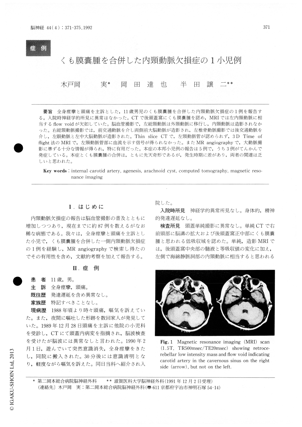
全身痙攣と頭痛を主訴とした,11歳男児のくも膜嚢腫を合併した内頸動脈欠損症の1例を報告する。入院時神経学的所見に異常はなかった。CTで後頭蓋窩にくも膜嚢腫を認め,MRIでは左内頸動脈に相当するnow oidが欠如していた。脳血管撮影で,左総頸動脈は外頸動脈に移行し,内頸動脈は造影されなかった。右総頸動脈撮影では,前交通動脈を介し両側前大脳動脈が造影され,左椎骨動脈撮影では後交通動脈を介し,左眼動脈と左中大脳動脈が造影された。Thin slice CTで,左頸動脈管が認められず,3D Time ofHight法のMRIで,左頸動脈管部に血流を示す信号が得られなかった。またMR angiographyで,大動脈撮影に準ずる十分な情報が得られ,特に有用だった。本症の本邦小児例の報告は5例で,うち3例がてんかんで発症している。本症とくも膜嚢腫の合併は,ともに先天奇形であるが,発生時期に差があり,両者の関連は乏しいと思われた。
A case of agenesis of the internal carotid artery combined with arachnoid cyst is reported.
This 11-year-old boy had occasionally com-plained headache and nausea since he was of 9 years old. He was admitted to our hospital because of an epileptic seizure. Physical and neurological exami-nations on admission were normal. A CT scan showed a cystic mass in retrocerebellar region. MRI suggested absence of flow void area indicating internal carotid artery in the cavernous sinus on left side.
Left common carotid angiogram showed absence of the internal carotid artery. Bilateral A2 seg-ments were supplied by right Al with tortuous anterior communicating artery. Left middle cere-bral artery and left ophthalmic artery were supplied via dilated left posterior communicating artery on left vertebral angiogram. Thin slice, axial target image of the CT revealed absence of the left bony carotid canal. MRI by 3D TOF method confirmed no blood flow in this area. MR angiography provided sufficient information about cervical vessels non-invasively. '"I-IMP SPECT image ascertained no hypoperfusion area in left cerebral hemisphere.
Convulsion was controlled with sodium valproate. Association of agenesis of the internal carotid artery and arachnoid cyst could be a coincidence.

Copyright © 1992, Igaku-Shoin Ltd. All rights reserved.


