Japanese
English
- 有料閲覧
- Abstract 文献概要
- 1ページ目 Look Inside
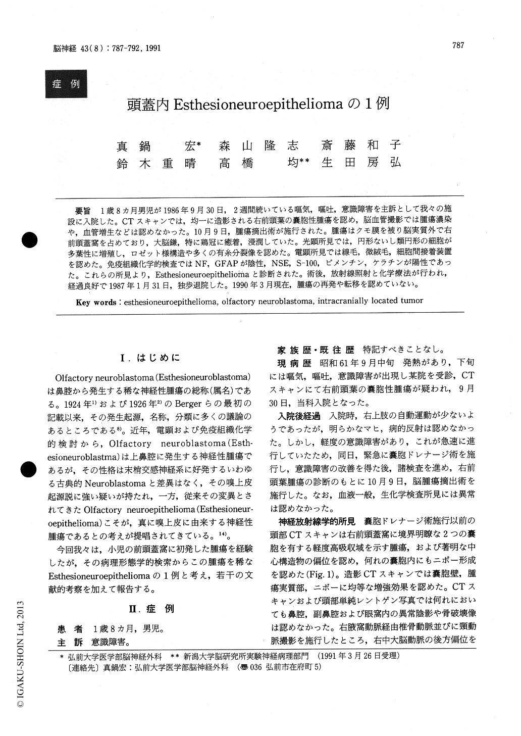
1歳8ヵ月男児が1986年9月30日,2週間続いている嘔気,嘔吐,意識障害を主訴として我々の施設に入院した。CTスキャンでは,均一に造影される右前頭葉の嚢胞性腫瘍を認め,脳血管撮影では腫瘍濃染や,血管増生などは認めなかった。10月9日,腫瘍摘出術が施行された。腫瘍はクモ膜を被り脳実質外で右前頭蓋窩を占めており,大脳鎌,特に鶏冠に癒着,浸潤していた。光顕所見では,円形ないし類円形の細胞が多葉性に増殖し,ロゼット様構造や多くの有糸分裂像を認めた。電顕所見では線毛,微絨毛,細胞間接着装置を認めた。免疫組織化学的検査ではNF,GFAPが陰性,NSE,S−100,ビメンチン,ケラチンが陽性であった。これらの所見より,Esthesioneuroepitheliomaと診断された。術後,放射線照射と化学療法が行われ,経過良好で1987年1月31日,独歩退院した。1990年3月現在,腫瘍の再発や転移を認めていない。
A 1-year-8-month-old boy was admitted to our service on September 30, 1986, complaining of nausea, vomiting and consciousness disturbance lasted for about 2 weeks. In CTs, right frontal cystic mass which was homogenously enhanced by contrast media was revealed. Neither hypervascularity nor tumor staining were seen agiographically. On October 9, 1986, total removal of the tumor was performed. The tumor waslocated extracerebrally in the right anterior cranial fossa, but was covered with arachnoid membrane. The tumor showed tight adhesion with falx cerebri, par-ticulary at crista galli where an invasive infiltration was seen. Light microscopic examination demonstrated oval or spherical small cells arranged multilobularly with rosette like formation and numerous mitoses. Ultras-tructurally, cilia, microvilli and junctional complexes were observed. No dense-cored secretory granules were found in the tumor cells. Immunohistochemical study onthis tumor showed negative NF and GFAP ; positive NSE, S-100, vimentin and keratin. From these findings, the tumor was diagnosed as esthesioneuroepithelioma. Postoperatively, irradiation and chemotherapies were also performed, and the patient showed uneventful course. On January 31, 1987, he was discharged on his foot, and no recurrent or metastatic signs could be found untill the end of March of 1990.

Copyright © 1991, Igaku-Shoin Ltd. All rights reserved.


