Japanese
English
- 有料閲覧
- Abstract 文献概要
- 1ページ目 Look Inside
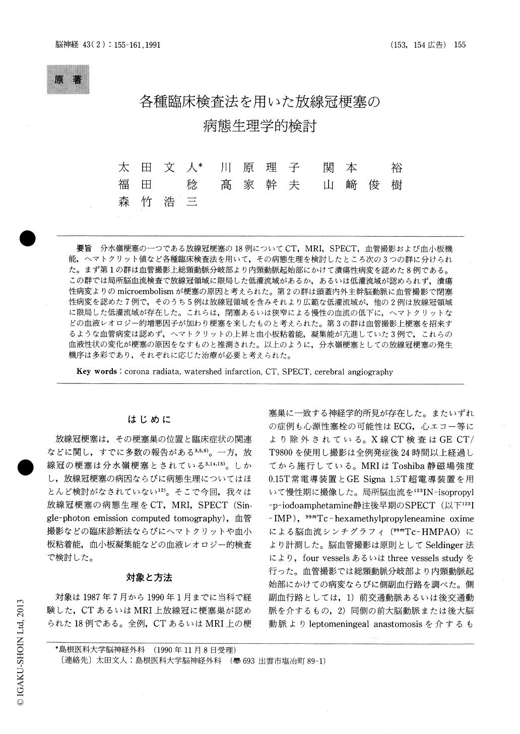
分水嶺梗塞の一つである放線冠梗塞の18例についてCT,MRI,SPECT,血管撮影および血小板機能,ヘマトクリット値など各種臨床検査法を用いて,その病態生理を検討したところ次の3つの群に分けられた。まず第1の群は血管撮影上総頸動脈分岐部より内頸動脈起始部にかけて潰瘍性病変を認めた8例である。この群では局所脳血流検査で放線冠領域に限局した低灌流域があるか,あるいは低灌流域が認められず,潰瘍性病変よりのmicroembolismが梗塞の原因と考えられた。第2の群は頭蓋内外主幹脳動脈に血管撮影で閉塞性病変を認めた7例で,そのうち5例は放線冠領域を含みそれより広範な低灌流域が,他の2例は放線冠領域に限局した低灌流域が存在した。これらは,閉塞あるいは狭窄による慢性の血流の低下に,ヘマトクリットなどの血液レオロジー的増悪因子が加わり梗塞を来したものと考えられた。第3の群は血管撮影上梗塞を招来するような血管病変は認めず,ヘマトクリットの上昇と血小板粘着能,凝集能が亢進していた3例で,これらの血液性状の変化が梗塞の原因をなすものと推測された。以上のように,分水嶺梗塞としての放線冠梗塞の発生機序は多彩であり,それぞれに応じた治療が必要と考えられた。
The authors evaluated 18 patients who presented with corona radiata infarction, one of the 'water-shed infarctions', on CT and/or MRI to determine its eitology and pathophysiology using cerebral angiography, single-photon emission computedtomography (SPECT), and tests of hemostatic func-tion including hematocrit, platelet aggregation and adhesiveness. On angiography, 8 of these 18 patients had ulcerative lesions in the common car-otid artery bifurcation with or without minimal stenosis and exhibited no or a minimal area of hypoperfusion localized to the corona radiata on SPECT. In these, microembolism from the lesions at the common carotid bifurcation seemed play an important role in the genesis of corona radiata infarction. In 7 of the remaining 10 patients, cere-bral angiography showed occlusive lesions of the internal carotid artery around its origin in 3, more than 90% stenosis of the internal carotid artery in 1, severe stenosis of the M1 segment of the middle cerebral artery in 2, and M1 occlusion in 1. In 5 of these 7, SPECT demonstrated a larger area of hypoperfusion than the corona radiata in the involved hemisphere. In the remaining 2, SPECT demonstrated a hypoperfusion area localized to the corona radiata. In all 7, the hematocrit was ele-vated. A collateral blood supply was visualized in 5 of 7 on cerebral angiography. In these 7 patients, hemodynamic disturbance was considered to con-tribute to the pathogenesis of infarction in the corona radiata. In the final three patients, cerebral angiography showed significant occlusive lesions in the main trunk of the cerebral arteries. In these 3 patients, no hypoperfusion area in the corona radiata was demonstrated on SPECT. All 3 patients had an enhancement of platelet aggregation and/or adhesiveness and an increase in the hematocrit. It appears that in these 3 cases, enhancement of aggre-gation and/or adhesiveness might have caused infarction in the corona radiata. There seem to be various mechanisms, such as microembolism from ulceration of atheromatous lesions in the common carotid bifurcation, hemodynamic disturbance as-sociated with occlusive lesions in the main trunk of the cerebral arteries, increase in hematocrit, and enhancement of platelet aggregation. And they have an important role in the pathogenesis of corona radiata infarction. Therefore, patients with corona radiata infarction must be treated in accor-dance with the pathogenesis of the lesion.

Copyright © 1991, Igaku-Shoin Ltd. All rights reserved.


