Japanese
English
- 有料閲覧
- Abstract 文献概要
- 1ページ目 Look Inside
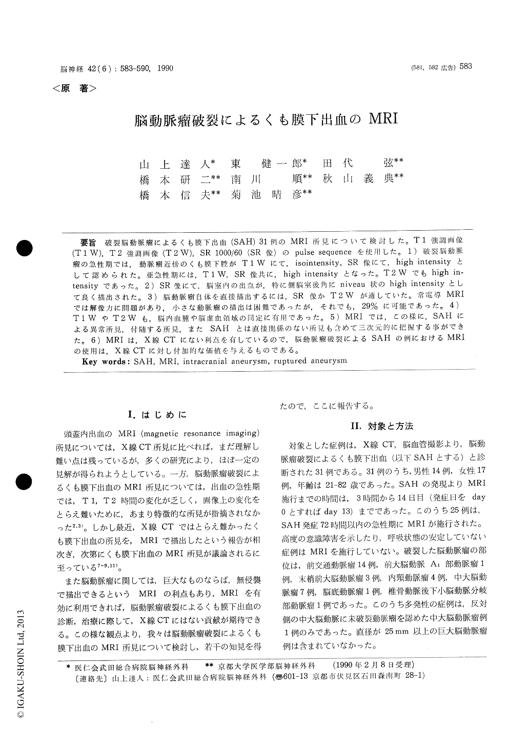
破裂脳動脈瘤によるくも膜下出血(SAH)31例のMRI所見について検討した。T1強調画像(T1W),T2強調画像(T2W),SR 1000/60(SR像)のpulse sequenceを使用した。1)破裂脳動脈瘤の急性期では,動脈瘤近傍のくも膜下腔がT1Wにて,isointensity, SR像にて,high intensityとして認められた。亜急性期には,T1W, SR像共に,high intensityとなった。T2Wでもhjgh in—tensityであった。2)SR像にて,脳室内の出血が,特に側脳室後角にniveau状のhigh intensityとして良く描出された。3)脳動脈瘤自体を直接描出するには,SR像かT2Wが適していた。常電導MRIでは解像力に問題があり,小さな動脈瘤の描出は困難であったが,それでも,29%に可能であった。4)T1WやT2Wも,脳内血腫や脳虚血領域の同定に有用であった。5)MRIでは,この様に,SAHによる異常所見,付随する所見,またSAHとは直接関係のない所見も含めて三次元的に把握する事ができた。6)MRIは,X線CTにない利点を有しているので,脳動脈瘤破裂によるSAHの例におけるMRIの使用は,X線CTに対し付加的な価値を与えるものである。
We report MRI (magnetic resonance imaging) findings of 31 cases with subarachnoid hemorrhage (SAH) due to ruptured intracranial aneurysm. Among these cases, 25 were studied within 72 hours after onset of SAH.
A resistive type MRI scanner operating at a field of 0. 2 Tesla was used in obtaining inversion recovery (IR) and saturation recovery (SR) images. IR 1500/43/400 (T1 weighted image,T1W), SR 1000/ 60 (SR image) and SR 2000/100 (T2 weighted image, T2W) were chosen for analysis. The SR image was usually adopted and coronal and/or sagittal images were added when there was enough time for examination. Slice thickness was 10 mm and slice interval was 15 mm. The scan was not necessarily aimed at the visualization of aneurysm itself.
1) In the acute phase of SAH, subarachnoidspaces near the ruptured aneurysm were appeared as isointensity areas on T1W and as high intensity areas on SR image. In the subacute phase, they were depicted as high intensity areas on both T1W and SR images, and as high intensity areas on T2W. 2) Intraventricular hemorrhage was visua-lized as a niveau-like high intensity area, especial-ly, within the posterior horns on the SR images. 3) SR and T2W images were suitable for detection of aneurysm itself. In a resistive-type scanner, small aneurysms were not easily visualized due to the lack of high resolution. However, 29% of aneuysms, which were not giant, could be visua-lized on MRI. 4) Identification of intracerebral hemorrhage and cerebral ischemia from various causes was easy on both T1W and T2W images. 5) By MRI, these abnormal findings due to SAH and other unrelated findings were understood as three-dimensional image. 6) MRI examination was useful in the cases with SAH due to ruptured intracranial aneurysms from the point of view that MRI is complementary to x-ray CT.

Copyright © 1990, Igaku-Shoin Ltd. All rights reserved.


