Japanese
English
- 有料閲覧
- Abstract 文献概要
- 1ページ目 Look Inside
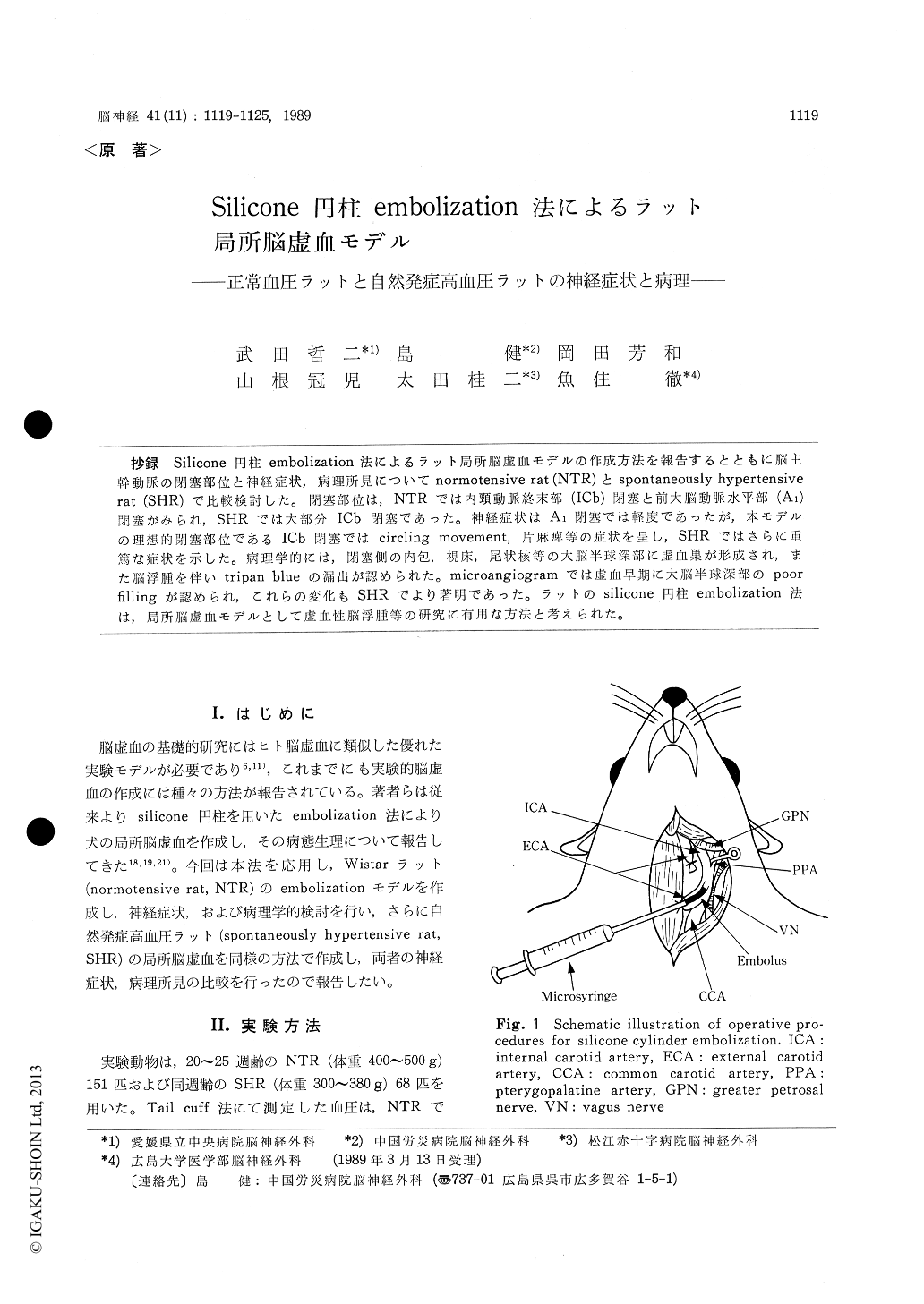
抄録 Silicone円柱embolization法によるラット局所脳虚血モデルの作成方法を報告するとともに脳主幹動脈の閉塞部位と神経症状,病理所見についてnormotensive rat (NTR〉とspontaneously hypertensiverat (SHR)で比較検討した。閉塞部位は,NTRでは内頸動脈終末部(ICb)閉塞と前大脳動脈水平部(A1)閉塞がみられ,SHRでは大部分ICb閉塞であつた。神経症状はA1閉蜜では軽度であったが,本モデルの理想的閉塞部位であるICb閉塞ではcircling movement,片麻痺等の症状を呈し,SHRではさらに重篤な症状を示した。病理学的には,閉塞側の内包,視床,尾状核等の大脳半球深部に虚血巣が形成され,また脳浮腫を伴いtripan blueの漏出が認められた。microangiogramでは虚血早期に大脳半球深部のpoorfillingが認められ,これらの変化もSHRでより著明であった。ラットのsilicone円柱embolization法は,局所脳虚血モデルとして虚血性脳浮腫等の研究に有用な方法と考えられた。
A focal cerebral ischemic model was produced by occlusion of the intracranial main cerebral artery with a silicone cylinder in normotensive (NTR) and spontaneously hypertensive rats (SHR). Main cerebral artery could be successfully occlu-ded in approximately 90%. The most frequent embolized site was the distal part of the internal cerebral artery (ICb) and less frequently the ho-rizontal segment of the anterior cerebral artery (Al). Mortality rate of NTR with ICb occlusion (NTR-ICb) was 43% at 72 hours after emboliza-tion and that of SHR with ICb occlusion (SHR-ICb) was 67% at 24 hours after embolization. NTR-ICb showed neurological signs (i. e. circling movement, hemiparesis, poor response to pain sti-muli) and histologically, showed infarction in the deep cerebral structures (i. e. thalamus, hypothala-mus, hippocampus, and internal capsule) accompa-nied with mild disruption of blood-brain barrier (BBB). SHR-ICb showed more serious neurological signs and more severe cerebral infarction in the deep cerebral structures with severe disruption of BBB.
In SHR-ICb, ischemic cerebral edema was more prominent which may deteriorated symptoms and pathological findings compared to NTR-ICb. This embolization model is proposed to be useful for studying the pathophysiology of focal cerebral ischemia, especially, early ischemic edema.

Copyright © 1989, Igaku-Shoin Ltd. All rights reserved.


