Japanese
English
- 有料閲覧
- Abstract 文献概要
- 1ページ目 Look Inside
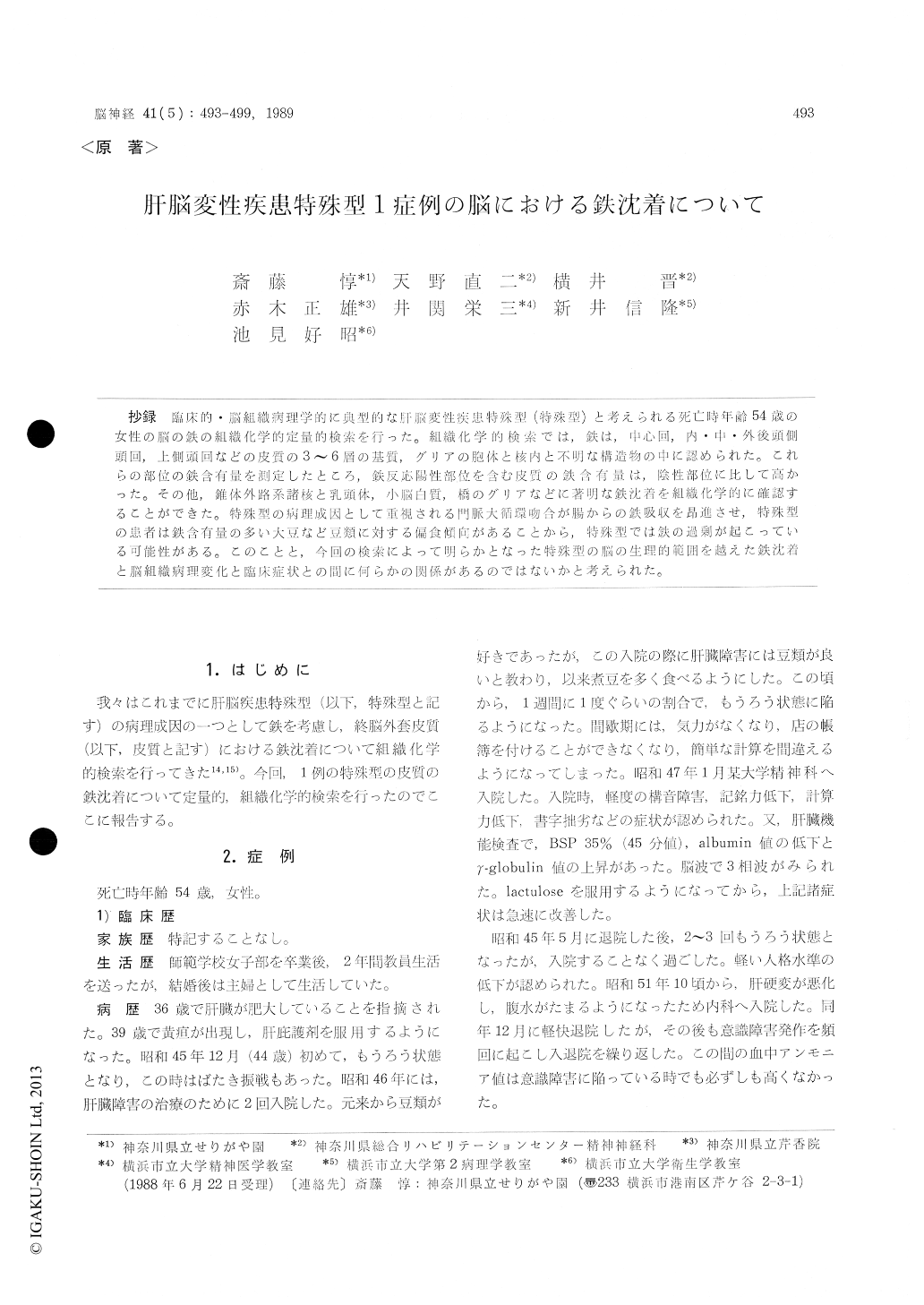
抄録 臨床的・脳組織病理学的典型的な肝脳変性疾患特殊型(特殊型)と考えられる死亡時年齢54歳の女性の脳の鉄の組織化学的定量的検索を行った。組織化学的検索では,鉄は,中心回,内・中・外後頭側頭回、上側頭回などの皮質の3〜6層の基質,グリアの胞体と核内と不明な構造物の中に認められた。これらの部位の鉄含有量を測定したところ,鉄反応陽性部位を含む皮質の鉄含有量は,陰性部位に比して高かった。その他,錐体外路系諸核と乳頭体,小脳白質,橋のグリアなどに著明な鉄沈着を組織化的に確認することができた。特珠型の病理成因として重視される門脈大循環吻合が腸からの鉄吸収を昂進させ,特殊型の患者は鉄含量の多い大豆など豆類に対する偏食傾向があることから,特殊型では鉄の過剰が起こっている可能性がある。このことと,今回の検索によって明らかとなった特殊型の脳の生理的範囲を越えた鉄沈着と脳組織病理変化と臨床症状との間に何らかの関係があるのではないかと考えられた。
The histochemical demonstration of iron and the iron content was examined in the brain of a case of the special type of hepatocerebral encephalo-pathy (I-ICE).
The patient had suffered from a liver disease since 36 years old. At 44 years old, she experi-enced the first attack of twilight state with flap-ping tremor. She had predilection for eating beans. Her personality gradually became euphoric with the recurrent episodes of unconsciousness. At 54 years old, she died of the complication of melena, renal insufficiency and pneumonia. The liver showed cirrhotic changes and iron con-tent of liver was 0 or 1 after MacDonald's criterion scale.
The histopathological findings of the brain showed the characteristic changes of HCE, which were incomplete softening and spongy state pseu-dolaminarilly extending in the deep layer of thecerebral cortex, the proliferation of the severely changed Alzheimer 2 type glias with or without intranuclear carmine positive substance.
The deparaffinized sections, 20 t in thickness, which were not fastened on slides were used for the histochemical study of iron, because iron depo-sits displaced inside of the brain tissues when the paraffine sections were fastened on slide glasses in the constant-temperature bath. The iron deposi-tion was found in the central gyrus, superior tem-poral gyrus, medial and lateral occipito-temporal gyrus and middle temporal gyrus of occipital lobe. The iron accumulated in the ground substance, glia cell bodies, glia nuclei and unknown bodies in the 3-6 layers of cerebral cortex of these gyri. The iron accumulation demonstrated histochemically inother parts of the brain were group 1, 2 by Spatz, mammillary body, glia cell bodies in cerebellar white matter and pons.
Biochemically iron content increased in iron po-sitive cerbral cortex.
In this study, iron content increased in the cere-bral cortex of HCE. The portacaval shunt which plays an important role in the pathogenesis of HCE increased iron absorption through the small intestine, while beans which often likes to be eaten by the patient of HCE is one of the foods including large amount of iron. By reason of these facts, it was considered that iron might have some clue to the pathogenesis of HCE.

Copyright © 1989, Igaku-Shoin Ltd. All rights reserved.


