Japanese
English
- 有料閲覧
- Abstract 文献概要
- 1ページ目 Look Inside
はじめに
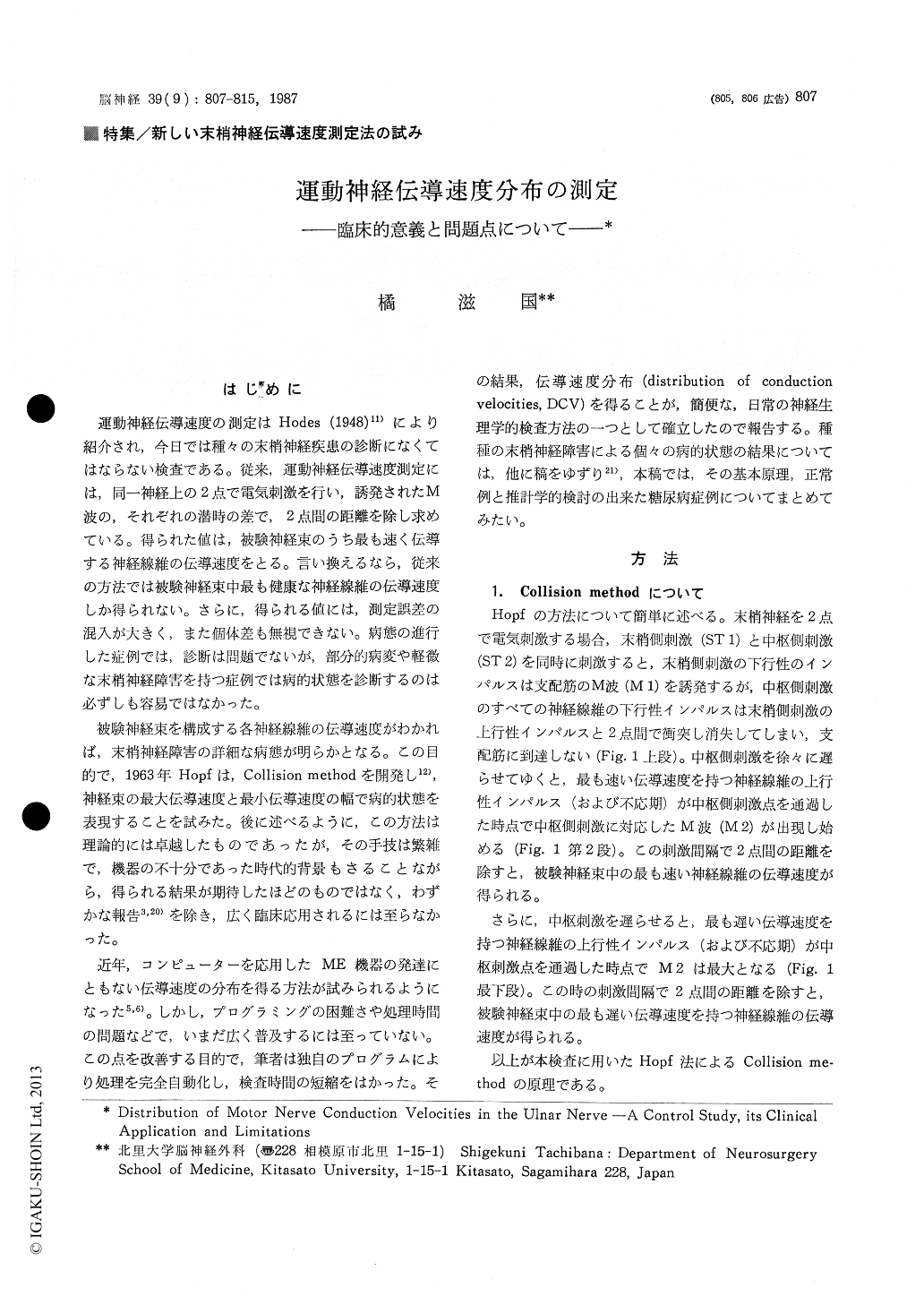
運動神経伝導速度の測定はHodes (1948)11)により紹介され,今日では種々の末梢神経疾患の診断になくてはならない検査である。従来,運動神経伝導速度測定には,同一神経上の2点で電気刺激を行い,誘発されたM波の,それぞれの潜時の差で,2点間の距離を除し求めている。得られた値は,被験神経束のうち最も速く伝導する神経線維の伝導速度をとる。言い換えるなら,従来の方法では被験神経東中最も健康な神経線維の伝導速度しか得られない。さらに,得られる値には,測定誤差の混入が大きく,また個体差も無視できない。病態の進行した症例では,診断は問題でないが,部分的病変や軽微な末梢神経障害を持つ症例では病的状態を診断するのは必ずしも容易ではなかった。
被験神経束を構成する各神経線維の伝導速度がわかれば,末梢神経障害の詳細な病態が明らかとなる。この目的で,1963年Hopfは,Collision methodを開発し12),神経束の最大伝導速度と最小伝導速度の幅で病的状態を表現することを試みた。後に述べるように,この方法は理論的には卓越したものであったが,その手技は繁雑で,機器の不十分であった時代的背景もさることながら,得られる結果が期待したほどのものではなく,わずかな報告3,20)を除き,広く臨床応用されるには至らなかった。
The goal of measuring the conduction velocity of a peripheral nerve is to describe the distri-bution of conduction velocities (DCV) of eachnerve fiber in a nerve bundle examined. The author devised a fully automatized method to describe the DCV of a nerve bundle with a signal processor. The principle of the method is nerve impulse collision method which was devised by Hopf in 1963. DCV of the ulnar nerve inner-vating the first dorsal interosseous muscle from the elbow to the wrist was examined in sixty five normal subjects without any evidence of pe-ripheral nerve problems, in ninety seven patients with diabetes mellitus (DM) and in five normal subjects whose forearm was cooled. Maximum conduction velocity at the same region was measured by using conventional method and com-pared to the DCV.
In control group, there was very mild influence of aging on the peak velocity of DCV but there was no statistically significant deference of mean DCV pattern between any decade of age. In this group a bimodal distribution of velocities was demonstrated. It is assumed that the distribution represents populations of motor fibers innervating the fast and the slow twitch muscle fibers in this muscle.
Those results in patients with DM and those normal subjects with their forearm was cooled were controversial. In patients with DM the peak velocity of DCV and its density has positive correlation, in contrast those cold injury group has negative correlation. It might be represents that the pathological condition of large fiber loss in the former group and small fiber damage in the latter group.
As it takes only five minutes to complete the whole examination, this method is available for use in routine clinical neurophysiological work.

Copyright © 1987, Igaku-Shoin Ltd. All rights reserved.


