Japanese
English
- 有料閲覧
- Abstract 文献概要
- 1ページ目 Look Inside
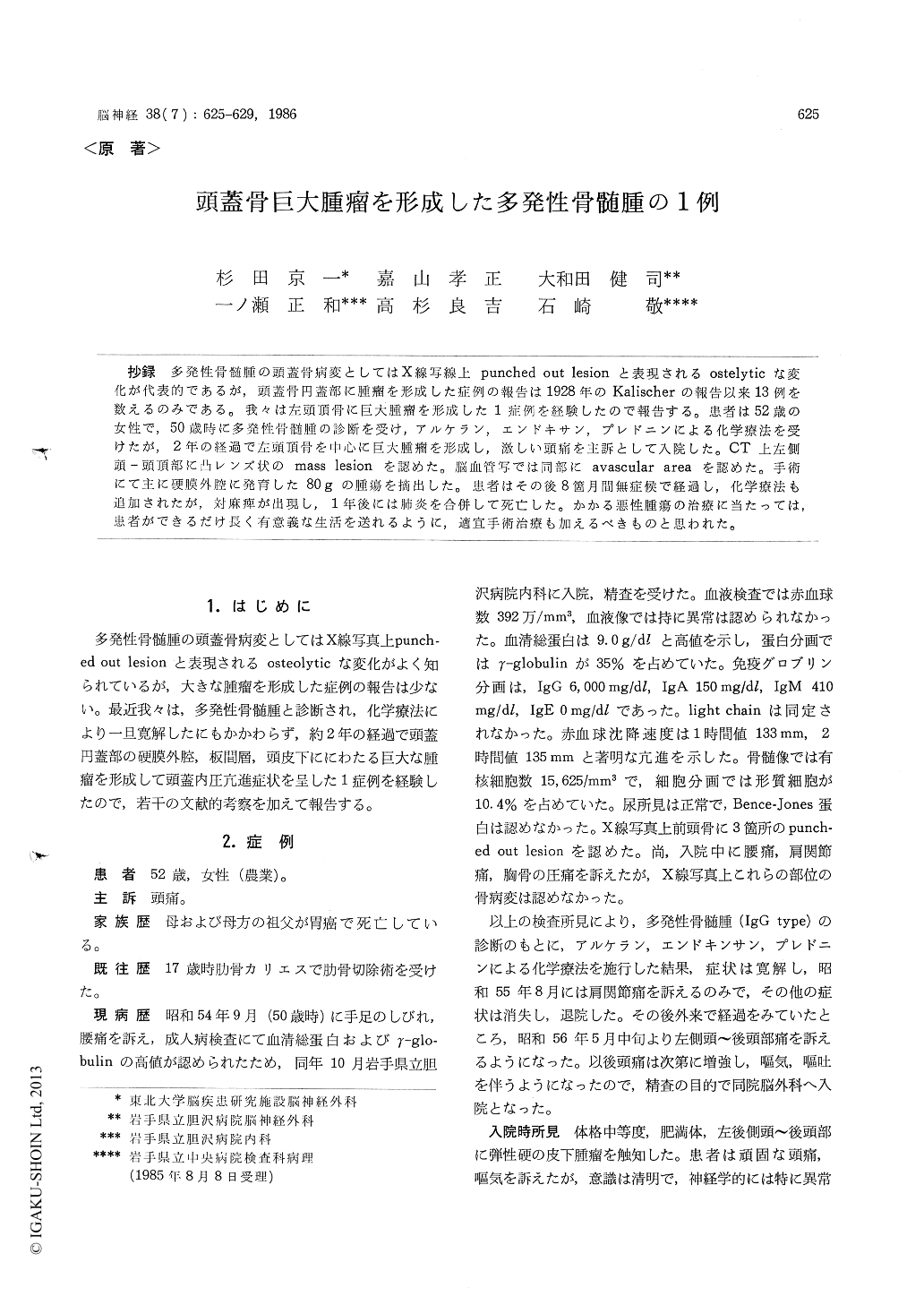
抄録 多発性骨髄腫の頭蓋骨病変としてはX線写線上punched out lesionと表現されるostelyticな変化が代表的であるが,頭蓋骨円蓋部に腫瘤を形成した症例の報告は1928年のKalischerの報告以来13例を数えるのみである。我々は左頭頂骨に巨大腫瘤を形成した1症例を経験したので報告する。患者は52歳の女性で,50歳時に多発性骨髄腫の診断を受け,アルケラン,エンドキサン,プレドニンによる化学療法を受けたが,2年の経過で左頭頂骨を中心に巨大腫瘤を形成し,激しい頭痛を主訴として入院した。CT上左側頭—頭頂部に凸レンズ状のmass lesionを認めた。脳血管写では同部にavascular areaを認めた。手術にて主に硬膜外腔に発育した80gの腫瘍を摘出した。患者はその後8箇月間無症候で経過し,化学療法も追加されたが,対麻痺が出現し,1年後には肺炎を合併して死亡した。かかる悪性腫瘍の治療に当たっては,患者ができるだけ長く有意義な生活を送れるように,適宜手術治療も加えるべきものと思われた。
It is well known that the case of multiple mye-loma shows punched-out lesions of the cranium without intracranial hypertension. In this paper a case of mulitple myeloma is reported showing intracranial hypertension due to a large tumor that developed in the left parietal bone. There are only 13 case reports about cranial mass lesion of multiple myeloma since 1928.
A 52 year-old female was admitted to Iwate Prefectural Isawa Hospital suffering from headache, nausea and vomiting. She had been already diag-nosed as multiple myeloma and treated with che-motherapy using Cyclophosphamide, Melphalan and Prednisolone for 2 years. On admission, a large subcutaneous mass was presented on the left parie-tal region. Craniogram revealed large osteolytic lesion of the left parietal bone and 3 punched-out lesions of the frontal bone. CT scan revealed a large mass lesion in the left epidural space, diploe and subcutandous space. Angiography showed avas-cular area. Brain scintigram showed diffuse hot area. Other skeletal bones showed no abormality. Laboratory examination revealed high concentra-tion of γ-globulin and high erythrocyte sedimen-tation rate. Electrophoresis showed high value of immunoglobulin G; immunoglobulin assay was as follows: IgG-6000mg/dl, IgA-150mg/dl, IgM-410mg/dl, IgE-0mg/dl. Serum electrolytes were with-in normal limits. Urine didn't include Bence-Jones protein. The patient was diagnosed as mul-tiple myeloma suffering from intracranial hyper-tension caused by large tumor which developed in the left parietal bone.
On the operation, large tumor was existed in the epidural and subcutaneous space invading into the diploe but without infiltration into the dura mater or cerebral cortex. The tumor was saucer-like and its diameter was 8cm. The tumor weigh-ed 80 gram. After the total removal of the tu-mor, she was free from any symptom and did well for 8 months after the surgery. But then she showed paraparesis and died due to pneumo-nia 1 year after the surgery.
In this case, large tumor developed in the skull in spite that the remission was achieved by che-motherapy using Cyclophosahamide, Melphalan and Prednisolon.

Copyright © 1986, Igaku-Shoin Ltd. All rights reserved.


