Japanese
English
- 有料閲覧
- Abstract 文献概要
- 1ページ目 Look Inside
抄録 てんかんの発生機序の解明に当り脳の各部位におけるてんかん発生の難易性を調べることは有意義なことと思われる。それ故作用機序が広く探求されているペニシリン(Pc)を用いて部位による難易性を調べた。当該部位以外からの影響を排除するために脳切片法を用いた。モルモットの海馬,大脳皮質および小脳の切片を電気刺激して誘発電位を起させ,それにPcを作用させててんかん様発作波が重なるかどうかを調べた。結果として,海馬が平均223u/mlで一番発作波が発生しやすく,次で大脳皮質520u/mlであるが,小脳では100〜20,000u/mlの如何なる濃度でも発生しなかった。Pcによってなぜ小脳で発作波が生じないかは明らかではないが,Pcの催てんかん作用に関与していると思われているGABAの局所分布に関しては,海馬,大脳皮質および小脳の間に大きな差はなく,PcによるGABA 作用の抑制のみでは発作波の発生機序は説明し難い。発作波が発生するためには反回性抑制機構が抑制され,それに伴って反同性興奮機構が促進されることが必要であるとする説が有力であるが,小脳にはこの反回性興奮機構がないと思われる。すなわちこの実験結果は発作波の発生に反回性興奮機構が重要な役割を果すことを示唆する。
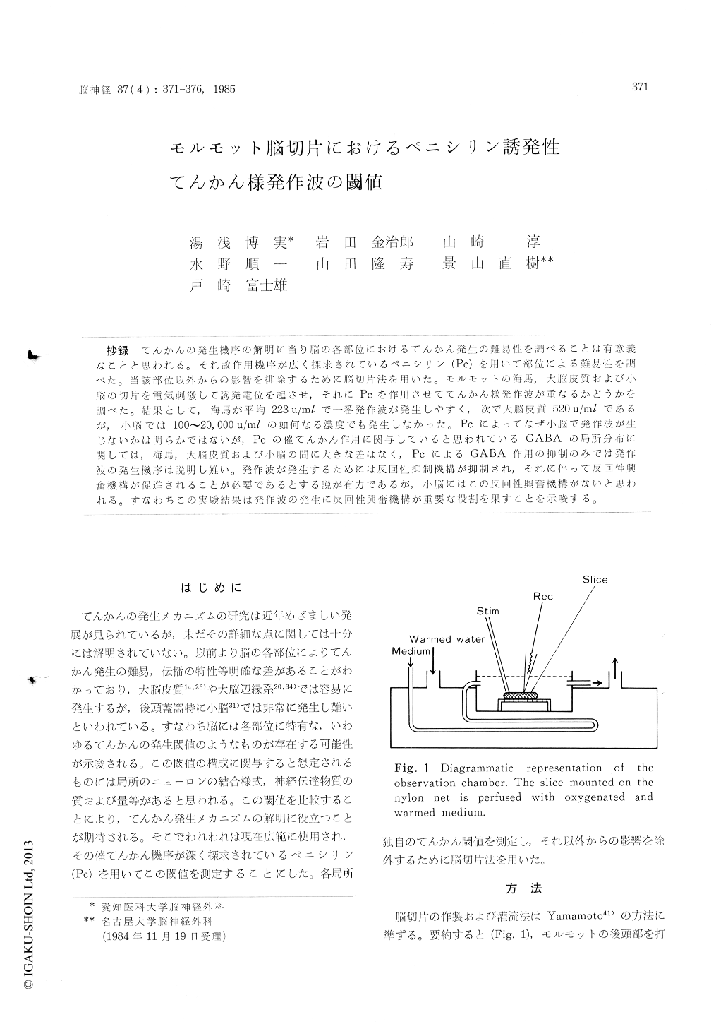
Investigation of the regional threshold for epi-lepsy in many structures in the brain would cont-ribute to the study of epileptogenesis. So we stu-died the threshold for epileptiform afterdischarge in the neocortex, hippocampus and cerebellar cortex using Na-Penicillin -G (Pc) of which epi-leptogenesis has been intensively investigated. In order to eliminate the influence from the outside of these structures, the brain slice method was utilized. Procedures for preparation of the tissue and incubation were about the same as those des-cribed by Yamamoto41). In summary, after sacrifice, brain of the guinea pig was taken out and the hippocampus, neocortex and cerebellum were cut with a razor blade under a microscope. The thick-ness of the section was about 0.3 mm. Slices were incubated at 37°C for about 30 min in the standard medium perfused with 95% 02 and 0% CO2. The chamber was continuously perfused with the standard medium which composed of NaCl, 124 mM, KC1, 5 : KH2P04, 1. 24 ; MgSO4, 1. 3 ; CaCl2, 2. 4 ; NaHCO3, 26 ; and glucose, 10.Evoked potentials were elicited by electrical sti-mulation in the standard medium. Mossy fibers were stimulated and responses were recorded from the CA 3 area in the hippocampus by glass pipette microelectrode. Subcortical white matter was sti-mulated and responses were recorded from the Purkinje cell layer in the cerebellum. Pc was add-ed in the standard medium until epileptiform afterdischarges were superimposed on the evoked potentials. The results of this experiment demons-trated that each structure in the brain has a re-gional own threshold for Pc induced epileptiform afterdischarge. Averaged threshold with 50% inci-dence (ED50) was 223 u/ml in the hippocampus, 520 u/ml in the neocortex, while no afterdischarge was elicited in the cerebellum even with the con-centration of 20, 000 u/ml.
The epileptogenesis of Pc is thought to result from two kinds of alteration in synaptic transmis-sion, i. e. increased excitation and decreased inhi-bition. Gamma-aminobutyric acid (GABA) would be an important transmitter in the epileptogenesis of Pc because Pc was reported to block inhibitory effect of GABA and also to inhibit GABA release from the nerve endings of the cortex. Okada et al29) reported that the regional distribution of GABA in guinea pig was as follows ; cortex cere-bellaris 2.48±0.33, cortex cerebralis 2.61 ±0.61, hippocampus 2,67±0.33 mmoles/kg. These figures are not in proportion to the threshold concentra-tion of Pc to elicit afterdischarge in this experi-ment, so it is postulated that epileptogenesis of Pc would not be explained only by suppression of GABA action.
It has been reported that in the neocortex and hippocampus epileptiform afterdischarge would be elicited by inhibition of recurrent inhibitory system and subsequent facilitation of recurrent excitatory system. It is well known, however, that the cerebellum does not possess this recur-rent excitatory system. The results of this expe-riment imply that the recurrent excitatory system plays a very important role in the epileptogenesis of Pc.

Copyright © 1985, Igaku-Shoin Ltd. All rights reserved.


