- 有料閲覧
- 文献概要
- 1ページ目
抄録 下垂体腺腫の分類において,電子顕微鏡の導入の意義は大きく,特にoncocytomaにおいては決定的であった。我々は,2例のoncocytomaを経験したので,臨床上の特徴と病理組織学上の特徴を述べ,あわせて本腫瘍の起源について考察した。本症は,中・高年者をに発生し,稀な例外を除いては非機能性腫瘍であり,従って来院時には大きな腫瘤を形成し,神経症状を呈している。CTスキャンでは,造影剤投与により強く増強される。細胞形態としては,他臓器のoncocytomaとは異なり,特別に大型とならず,染色性においてはミトコンドリアが染まるためエオジン好性を呈するが,アゾカルミンには染色されないため,嫌色素性・酸好性・塩基好性の三つに分類する古典的分類では,その本態を正確には伝えられないといえる。すなわち,本症の診断には電子顕微鏡が不可欠であることを述べた。本腫瘍の起源としては,下垂体自体の細胞であると考える。
Since the first reports of pituitary oncocytoma by Kovacs and Horvath, and Landolt and Oswald increasing numbers of cases have been reported with the advent of electron microscopy. It has been posturated that more cases were not precisely diagnosed because of lack of electron microscopic study.
We reported two cases of pituitary oncocytoma and discussed clinicopathological aspects.
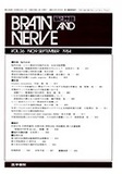
Case 1 A 66-year-old woman had a 6 year his-tory of visual impairment. Neurological examina-tion revealed loss of vision on the left and decreased visual acuity (0.3) with temporal hemianopsia on the right. The endocrinological study revealed moderate panhypopituitarism. Plain skull X-ray, computed tomography and cerebral angiography showed the findings of a pituitary tumor with suprasellar extension. Subfrontal removal of the tumor followed by irradiation was performed.
Case 2 A 50-year-old man was well until 8 years previously, when he experienced loss of libido. Three years before entry, the left sided exoph-thalmos and ptosis were noted. Neurological exa-mination showed a severe visual impairment witha bitemporal held detect, bilateral optic atrophy and disturbance of eye movements on the left. Endocrinological study revealed panhypopituita-rism. Radiological studies showed a pituitary tumor with a suprasellar extension and an invasion into the left orbital cavity. Transcranial and then transsphenoidal partial removal of the tumor were done followed by irradiation.
Histological examination of the tumors revealed a poorly granurated adenoma with very weak affinity to acid dyes. The tumor invaded into the orbital cavity in case 2 was similar to one in the cranial cavity. Immunohistochemical stains for growth hormone and prolactin were negative inboth cases. Electron microscopy revealed that both tumors were composed predominantly of typical oncocytes with abundant mitochondria of various shape and size. Few secretory granules ranging from 80 nm to 250nm in diameter were demonstrated.
Pituitary oncocytoma may be diagnosed by light microscopy if one recognizes exisistence of this type of tumor but precise diagnosis of the tumor is done by electron microscopy.
On the basis of the existence of secretory gran-ules and a wide variety in number of mitochondria in tumor cells, we posturate the oncocyte may be pituitary origin.
Copyright © 1984, Igaku-Shoin Ltd. All rights reserved.