Japanese
English
- 有料閲覧
- Abstract 文献概要
- 1ページ目 Look Inside
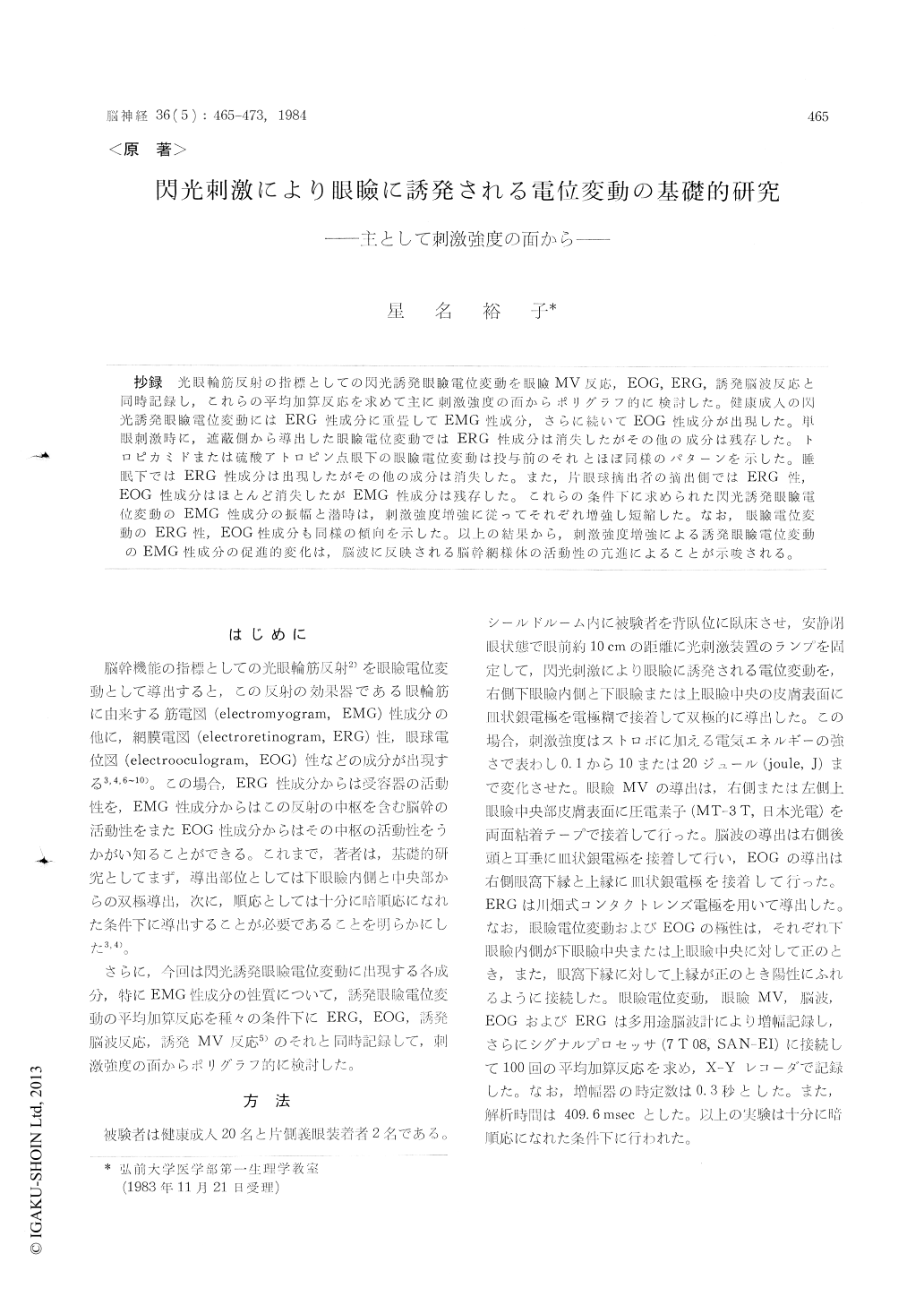
抄録 光眼輪筋反射の指標としての閃光誘発眼瞼電位変動を眼瞼MV反応,EOG,ERG,誘発脳波反応と当時記録し,これらの平均加算反応を求めて主に刺激強度の面からポリグラフ的に検討した。健康成人の閃光誘発眼瞼電位変動にはERG性成分に重畳してEMG性成分,さらに続いてEOG性成分が出現した。単眼刺激時に,遮蔽側から導出した眼瞼電位変動ではERG性成分は消失したがその他の成分は残存した。トロピカミドまたは硫酸アトロピン点眼下の眼瞼電位変動は投与前のそれとほぼ同様のパターンを示した。睡眠下ではERG性成分は出現したがその他の成分は消失した。また,片眼球摘出者の摘出側ではERG性,EOG性成分はほとんど消失したがEMG性成分は残存した。これらの条件下に求められた閃光誘発眼瞼電位変動のEMG性成分の振幅潜時は,刺激強度増強に従ってそれぞれ増強し短縮した。なお,眼瞼電位変動のERG性,EOG性成分も同様の傾向を示した。以上の結果から,刺激強度増強による誘発眼瞼電位変動のEMG性成分の促進的変化は,脳波に反映される脳幹網様体の活動性の亢進によることが示唆される。
The average photically evoked lid potential chan-ges are known to consist of electromyographic components due to the orbicularis oculi reflex, electroretinographic and electrooculographic com-ponents, which corresponded to the average sum-mating responses of the lid MV responses, elec-troretinogram (ERG), electrooculogram (EOG), respectively. In the present study the photically evoked lid potential changes were studied from the standpoint of the stimulus strength in healthy subjects and patients with an artificial eyeball. The photically evoked lid potential changes were obtained with the summation technique under vari-ous physiologic and pathologic conditions. In ad-dition, the average summating responses of the lid MV responses, EOG, ERG and EEG were also recorded simultaneously and discussed polygraphic-ally.
The average evoked lid potential changes in healthy resting subject with eyes closed were shown to be composed of the electromyographic components dependent on the effector activity of the orbicularis oculi reflex, early and late slow components which corresponded to the ERG and the EOG respectively.
The threshold stimulus necessary to elicit the electroretinographic, electromyographic and elec-trooculographic components was the intensity of flash stimulation, that is about 0. 1, 0. 3 and 4 joule in energy delivered to the stroboscope respectively. These components were observed to be increased in amplitude gradually according to the intensity strengthened.
In the average lid potential changes obtained by random flash stimulation to a single eye, the electroretinographic components in the occluded side disappeared completely, but the other com-ponents were clearly observed and gradually in-creased in amplitude according to the intensitystrengthened, although they were considerably decreased in amplitude than those obtained in the contralateral side of normal subjects.
After dropping tropicamide or atropine sulfate in the right eye, the patterns of lid potential changes were almost unchanged and increased in amplitude according to the intensity strengthened, though they are somewhat increased in amplitude and shortened in latent period.
In the average lid potential changes of a patient with an artificial eyeball, the electroretinographic components disappeared and the electrooculogra-phic components almost disappeared in the side of artificial one. But electromyographic compo-nents of evoked lid potential changes in the side of healthy and artificial eyeballs were increased in amplitude according to the intensity strength-ened, respectively, although they were consider-ably decreased in amplitude in the side of an arti-ficial eyeball.
In the process of natural sleep, the electromyo-graphic and electrooculographic components of the average evoked lid potential changes almost disap-peared in stage 2. The electroretinographic com-ponents, however, remained and were considerably increased in amplitude according to the intensity strengthened.
From the above results, it seems likely that the augmentative effect of the stimulus intensity strengthened on the average photically evoked lid potential changes may be produced by the increased activity of the facial nerves and the orbicularis oculi, through increased impulses that arrive at the reflex center of the brain stem from the retina, that is the receptor in the blinking reflex. In addition, it was suggested that the electrooculographic com-ponents of the evoked lid potential changes may also reflect the activities of brain stem related to eye movement.

Copyright © 1984, Igaku-Shoin Ltd. All rights reserved.


