Japanese
English
- 有料閲覧
- Abstract 文献概要
- 1ページ目 Look Inside
はじめに
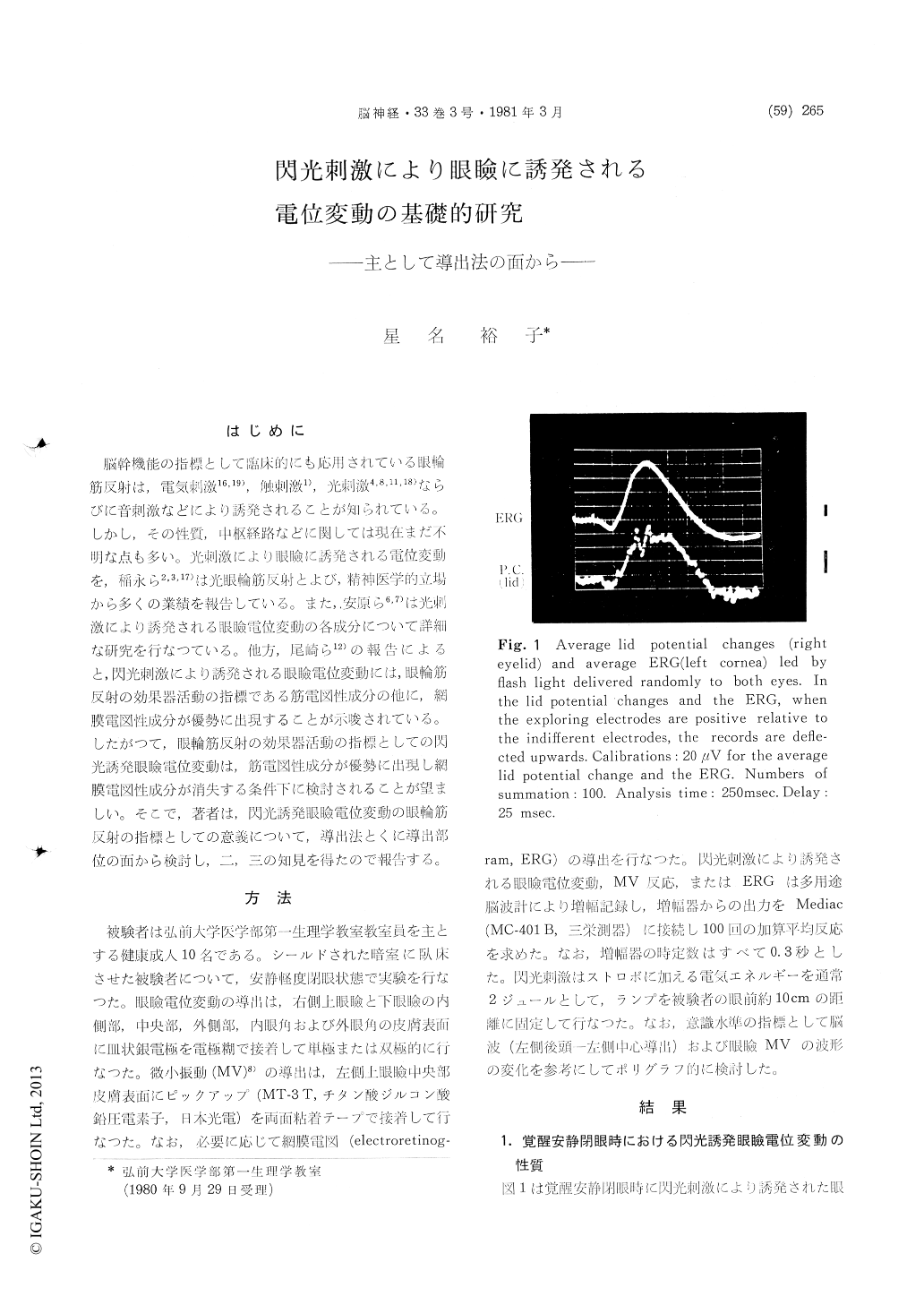
脳幹機能の指標として臨床的にも応用されている眼輪筋反射は,電気刺激16,19),触刺激1),光刺激4,8,11,18)ならびに音刺激などにより誘発されることが知られている。しかし,その性質,中枢経路などに関しては現在まだ不明な点も多い。光刺激により眼瞼に誘発される電位変動を,稲永ら2,3,17)は光眼輪筋反射とよび,精神医学的立場から多くの業績を報告している。また,安原ら6,7)は光刺激により誘発される眼瞼電位変動の各成分について詳細な研究を行なつている。他方,尾崎ら12)の報告によると,閃光刺激により誘発される眼瞼電位変動には,眼輪筋反射の効果器活動の指標である筋電図性成分の他に,網膜電図性成分が優勢に出現することが示唆されている。したがつて,眼輪筋反射の効果器活動の指標としての閃光誘発眼瞼電位変動は,筋電図性成分が優勢に出現し網膜電図性成分が消失する条件下に検討されることが望ましい。そこで,著者は,閃光誘発眼瞼電位変動の眼輪筋反射の指標としての意義について,導出法とくに導出部位の面から検討し,二,三の知見を得たので報告する。
In order to determine the most adequate method to lead the photically evoked lid potential changes, their physiological properties were studied from standpoint of the effector activity of the orbicularis oculi reflex. The photically evoked lid potential changes were obtained with the summation tech-nique in healthy subjects. In addition, the average responses of the photically evoked lid MV responses or electroretinograms were also recorded simultane-ously and discussed polygraphically. The following results were obtained.
1. In the average photically evoked lid potential changes led off unipolarly, the most adequate partof the indifferent electrode was found out to be the lobule, the exploring electrode being placed on the middle part of the superior lid.
2. In the average evoked lid potential changes led off unipolarly by placing one indifferent electrode on the ipsilateral lobule and the other exploring electrodes on different parts of the eyelid, the electromyographic components of the lid potential changes were observed to be the highest in ampli-tude, the exploring electrodes being placed on the medial parts of superior and inferior lids. However, the electroretinographic components in addition to electromyographic one were also shown to appear markedly in these cases.
3. In the average lid potential changes obtained by flash stimulation to a single eye, the electro-retinographic components disappeared almost com-pletely in the occluded side. On the other hand, the electromyographic components were observed to be more dominant in the medial parts of superior or inferior lid and medial angle than in the other parts of eyelid.
4. In the average lid potential changes led off bipolarly by exploring electrodes from different parts of the superior and inferior lids, the electro-retinographic components were shown to decrease considerably in amplitude as compared with those obtained by unipolar leads.
5. In the average lid potential changes led off bipolarly from middle and medial parts of the superior and inferior lids, the electromyographic components appeared markedly, although the elec-troretinographic components disappeared almost completely.
6. From these results, it seems to be likely that the bipolar leads from middle and medial parts of the superior or inferior lid may be the most ade-quate in order to obtain the average photically evoked lid potential changes.

Copyright © 1981, Igaku-Shoin Ltd. All rights reserved.


