Japanese
English
- 有料閲覧
- Abstract 文献概要
- 1ページ目 Look Inside
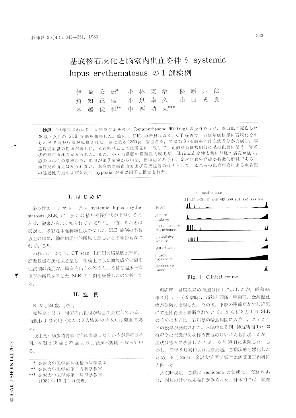
抄録 10年間にわたり,副腎皮質ホルモン(betamethasone 6000mg)の投与をうけ,脳出血で死亡した28歳・女性のSLE症例を報告した。臨床上DICの所見はなく,CT険査で,両側基底核部に石灰化をおもわせる高吸収域が観察された。脳は重さ1350g。脳室各部,特に第3・4脳室には血液成分が充満し,脳室周囲組織の崩が著しい。光顕所見としては所見に一致して,両側淡蒼球吻側部に毛細血管に沿う,顆粒状の類石灰沈着がみられた。また,小・細動脈の増殖性内膜肥厚,fibrinoid変性と共に静脈の病変が強く,静脈中心性の漿液浸潤,出血が第3脳室から中的脳,橋中心にみられ,2次的脳室穿破が特微的所見である。血管炎の所見はみられない。本症例の脳出血および石灰沈着の成因として,これらの血管病変による血管壁の透過性亢進および2次的hypoxiaが重要町因子と推測された。
A case of systemic lupus erythematosus with calcification of basal ganglia and intraventricular bleeding, was reported.
The patient, a 28-year-old woman was treated with steroid hormone therapy (total amount of steroid hormone ; betamethasone 6000 mg) during the years since the onset of her illness. At the end stage, she had a paresthesia in the lower extremities and suffered from high grade fever,and suddenly died of cerebral bleeding. Labola-tory findings showed no evidence of disseminated intravascular coagulopathy. A high density area in the bilateral pallidal parts was detected in the cerebral computed tomography.
General autopsy was performed.
Brain weight is 1350 g. Macroscopically, lateral, third, and forth ventricles are filled with massive blood and are dilatated with the destruction of periventricular tissues. Uncal and tonsillar hernia-tion are found. Microscopically, there are two important findings :
1) Bilateral rostral pallidum with little neuro-nal cell changes has calcification which is granular appearance along the capillary vessels or spicular like structure.
2) Most characteristic change is a perivascular, above all, perivenous transfusion, and there are ring like hemorrhage and plasma infiltration inthe saccular dilatated subadventitial space.
Thickening and fibrinoid degeneration of the vascular wall are also found, but there are no findings of vasculitis.
The degeneration of small arteries has been the most common pathological feature of the cerebral lesion in systemic lupus erythematosus, however our case has not only arterial lesions but also severe degeneration of venous wall, which may cause the bleeding or infarction.
This venous lesions are considered to be the important pathogenesis of the cerebral lesion in systemic lupus erythematosus, and these vascular lesion which may change the permeability of the blood vessel and cause the local hypoxia, is con-tributed to the calcification of the blood vessel and cause the local hypoxia, is attributable to the calcification of the pallidum.
Copyright © 1983, Igaku-Shoin Ltd. All rights reserved.