Japanese
English
- 有料閲覧
- Abstract 文献概要
- 1ページ目 Look Inside
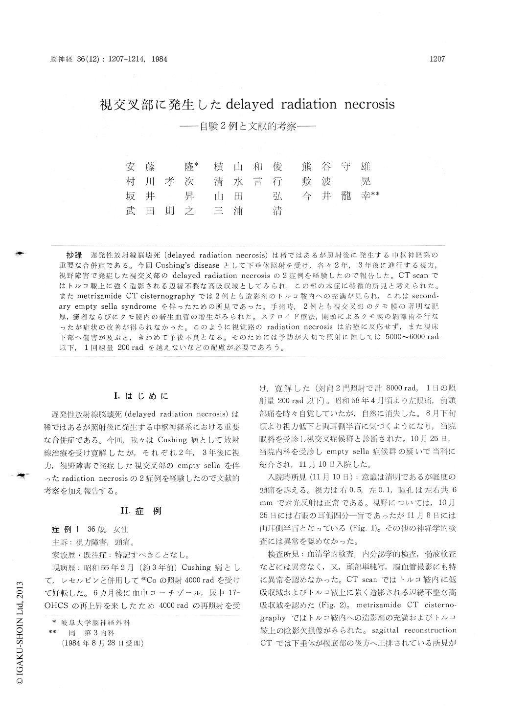
抄録 遅発性放射線脳壊死(delayed radiation necrosis)は稀ではあるが照射後に発生する中枢神経系の重要な合併症である。今回Cushing’s diseaseとして下垂体照射を受け,各々2年,3年後に進行する視力,視野障害で発症した視交叉部のdelayed radiation necrosisの2症例を経験したので報告した。CT scanではトルコ鞍上に強く造影される辺縁不整な高吸収域としてみられ,この部の本症に特徴的所見と考えられた。またmetrizamide CT cisternographyでは2例とも造影剤のトルコ鞍内への充満が見られ,これはsecond—ary empty sella syndromeを伴ったための所見であった。手術時,2例とも視交叉部のクモ膜の著明な肥厚,癒着ならぴにクモ膜内の新生血管の増生がみられた。ステロイド療法,開頭によるクモ膜の剥離術を行なったが症状の改善が得られなかった。このように視覚路のradiation necrosisは治療に反応せず,また視床下部へ傷害が及ぶと,きわめて予後不良となる。そのためには予防が大切で照射に際しては5000〜6000rad以下,1回線量200radを越えないなどの配慮が必要であろう。
Two cases with delayed radiation necrosis of the chiasmatic region following irradiation of the hypophysis for treatment of Cushing's disease were presented. Case 1 was a 36-year-old female who had reduction of visual acuity and bitemporal hemianopsia 2 years after 60Co-irradiation therapy (total 8000 rads) for Cushing's disease. CT scans showed low density in the pituitary fossa and ir-regular contrast-enhanced suprasellar mass, and metrizamide CT cisternography revealed the pit-uitary fossa filled with contrast medium. From those findings, secondary empty sella syndrome was suspicious. Case 2 was a 35-year-old male who had progressive visual disturbance 3 years after 60Co-irradiation therapy (total 9050 rads) for Cush-ing's disease. The right visual acuity was 0.05 and the left one was 0.1. Examination of visual field showed left homonymous hemianopsia. CT scans showed the contrast enhanced suprasellar mass extending to the right anterior thalamic region, and metrizamide CT cisternography de-tected secondary empty sella as same as that of Case 1.
Both cases were treated by administration of steroid hormone intrathecally and orally which was not effective. Right frontotemporal cranioto-mies were performed. Intraoperative findings of the chiasmatic region revealed diffuse adhesions and thickening of arachnoid membrane with mesh-work of fine vascular proliferations and adhesioto-mies were performed in both cases. On the post-operative course, visual disturbance did not im-proved in both cases. Histological examinations of the thicked arachnoids obtained intraoperatively showed the fibrous connective tissues with round cells infiltrations in both cases, and in Case 2, the small vessel in the arachnoid was occluded by organized thrombi.
Authors reviewed and analyzed literatures of delayed radiation necrosis. The incidence of this condition was 4% to 9% and onset of the symp-toms occured approximately 2 years after irradia-tion to hypophysis. Administration of steroid hor-mone and surgical treatment for the radiation necrosis involving the chiasmatic region were almost ineffective and also the prognosis of radio-necrotic lesions involving the hypothalamus was very poor. Therefore, radiotherapy for hypophy-seal region must be carried out by means of a rotation or arching technique in order to avoid this condition and further total dosage and its fractionation in radiation therapy should not exceed 6000 rads and 200 rads a day.

Copyright © 1984, Igaku-Shoin Ltd. All rights reserved.


