Japanese
English
- 有料閲覧
- Abstract 文献概要
- 1ページ目 Look Inside
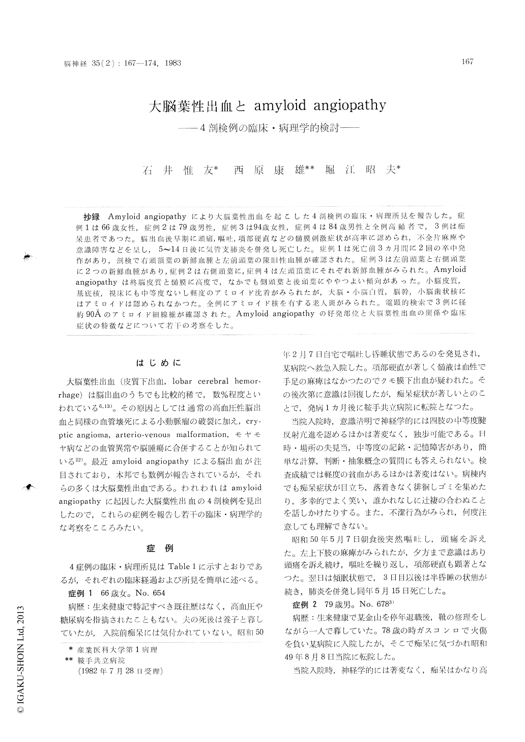
抄録 Amyloid angiopathyにより大脳葉性出血を起こした4剖検例の臨床・病理所見を報告した。症例1は66歳女性,症例2は79歳男性,症例3は94歳女性,症例4は84歳男性と全例高齢者で,3例は痴呆患者であつた。脳出血後早期に頭痛,嘔吐,項部硬直などの髄膜刺激症状が高率に認められ,不全片麻痺や意識障害などを呈し,5〜14日後に気管支肺炎を併発し死亡した。症例1は死亡前3カ月間に2回の卒中発作があり,剖検で右頭頂葉の新鮮血腫と左前頭葉の陳旧性血腫が確認された。症例3は左前頭葉と右側頭葉に2つの新鮮血腫があり,症例2は右側頭葉に,症例4は左頭頂葉にそれぞれ新鮮血腫がみられた。Amyloidangiopathyは終脳皮質と髄膜に高度で,なかでも側頭葉と後頭葉にややつよい傾向があった。小脳皮質,基底核,視床にも中等度ないし軽度のアミロイド沈着がみられたが,大脳・小脳白質,脳幹,小脳歯状核にはアミロイドは認められなかつた。全例にアミロイド核を有する老人斑がみられた、,電顕的検索で3例に径約90Åのアミロイド細線維が確認された。Amyloid angiopathyの好発部位と大脳葉性出血の関係や臨床症状の特徴などについて若干の考察をした。
Four autopsied cases of lobar cerebral hemorr-hage due to amyloid angiopathy were reported. They were 66 year old demented woman (Case 1), 79 year old man with typical clinical features of Amzheimer type senile dementia (Case 2), 94 year old slightly demented woman (Case 3) and 85 year old non-demented man (Case 4). There was no hypertension in these 4 patients, although Case 4 showed left ventricular hypertrophy at autopsy, suggestive of an unnoticed clinical hypertension. At the onset of stroke, there was a high incidence of meningeal irritation, such as headache, vomit-ting and nuchal rigidity They developed distur-bance of consciousness of various degree andhemiparesis. They expired 5 to 14 days after the onset, due to an associated bronchopneumonia. Case 1 had two episodes of cerebral hemorrhage within the last 3 months.An autopsy revealed a recent hematoma in the right parietal lobe and an old hematoma in the left frontal lobe. Case 3 had two recent hematomas in the left frontal and the right temporal lobes. Case 2 and 4 had single hematoma, in the right temporal and the left parietal lobes, respectively. All the hematomas were subcortical, lobar hemorrhages without involving the basal ganglia and the thalamus.
Microscopically the most striking finding was amyloid deposit in the wall of small arteries and arterioles of the cerebral cortex and the overlying leptomeninges (congophilic angio-pathy). Its intensity varied in the different parts of the central nervous system. The cerebral cor-tex was affected most severely in all cases, with some predilection of the occipital and temporal lobes. The cerebellar cortex, Ammon's horn, ba-sal ganglia, amygdala and thalamus exhibitted sli-ght to moderate amyloid deposit. There was no congophilic angiopathy in the white matter, ex-cept for Case 2, in which slight amyloid deposit was observed in the vessels of subcortical U-fiber. The brain stem and the dentate nucleus of the cerebellum showed no amyloid. Also present was prominent "drusige Entartung", namely amyloid deposit in the capillary wall with infiltration into the adjacent brain parenchyma. It was prominent in Case 2, moderate in Case 3 and slight in Case 1 and 4. The occipital and the temporal lobes were afftcted predilectively. Senile plaques with amyloid core were noted in all cases. There se-ems to be correlation between the presence of senile plaque and the congophilic angiopathy. Alzheimer's neurofibrillary tangle was also pre-sent in all cases, however in Case 4, it was limit-ted to Ammon's horn. Case 3 and 4 showed fibri-noid degeneration (angionecrosis) of the small arteries in the basal ganglia as well. There was no cryptic angioma in and around the hematomas.
Amyloid angiopathy is said to be most prominent in the cerebral cortex, particularly the temporal and occipital lobes. Therefore, its cerebral hemor-rhage is usually a lobar type, as compared to the common location of hypertensive hemorrhage, that is basal ganglia, thalamus, brain stem and dentate nucleus of the cerebellum. Amyloid angiopathy shold be considered as a source of lobar cerebral hemorrhage, if it occurs in the elderly, normo-tensive and demented patient.

Copyright © 1983, Igaku-Shoin Ltd. All rights reserved.


