Japanese
English
- 有料閲覧
- Abstract 文献概要
- 1ページ目 Look Inside
はじめに
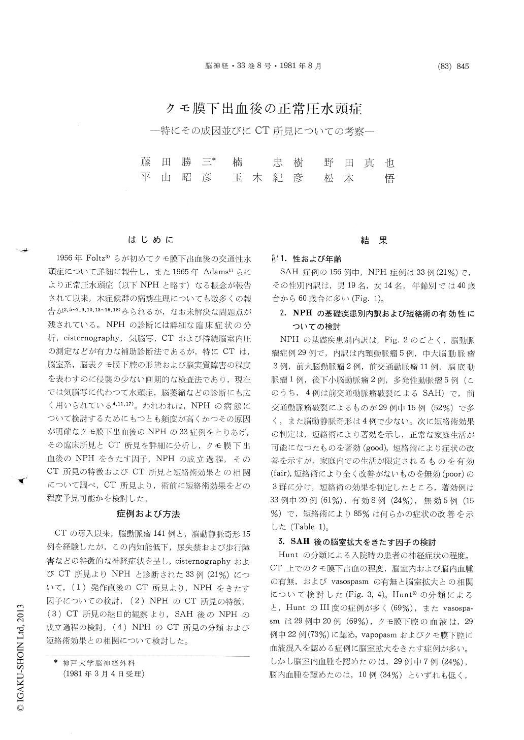
1956年Foltz3)らが初めてクモ膜下出血後の交通性水頭症について詳細に報告し,また1965年Adams1)らにより正常圧水頭症(以下NPHと略す)なる概念が報告されて以来,本症候群の病態生理についても数多くの報告が2,5〜7,9,10,13〜16,18)みられるが,なお未解決な問題点が残されている。NPHの診断には詳細な臨床症状の分析,cisternography,気脳写,CTおよび持続脳室内圧の測定などが有力な補助診断法であるが,特にCTは,脳室系,脳表クモ膜下腔の形態および脳実質障害の程度を表わすのに侵襲の少ない画期的な検査法であり,現在では気脳写に代わつて水頭症,脳萎縮などの診断にも広く用いられている4,11,17)。われわれは,NPHの病態について検討するためにもつとも頻度が高くかつその原因が明確なクモ膜下出血後のNPHの33症例をとりあげ,その臨床所見とCT所見を詳細に分析し,クモ膜下出血後のNPHをきたす因子,NPHの成立過程,そのCT所見の特徴およびCT所見と短絡術効果との相関について調べ,CT所見より,術前に短絡術効果をどの程度予見可能かを検討した。
CT scans were used to evaluate the development of the hydrocephalus, periventricular hypodensity (PVH) and the degree of the brain damage on 33 patients with the normal pressure hydrocephalus (NPH), following the subarachnoid hemorrhage (SAH), and the following conclusions can be drawn from our study.
1) NPH occurs in 33 cases out of 156 cases of subarachnoid hemorrhage (21%), and there was a relatively high incidence of normal pressure hydro-cephalus following rupture of the anterior com-municating artery aneurysm.
2) Factors causing NPH may be the arterial vasospasms and subarachnoid blood clot of the basal cistern verified by CT.
3) According to the repeated CT and lumbar tap after SAH, PVH, and, hydrocephalus usually be-come apparent around 7-10 days and most promi-nent around 3-4 weeks after SAH except for acute hydrocephalus appeared immediately after severe SAH.
4) The results were compared with CT findings and clinical response to shunting. The clinical improvements were achieved in cases (85%), inwhich CT showed PVH, small brain damage in the frontal lobe due to vasospasms or intracerebral hematoma, and no cortical atrophy.
5) Repeated CT can give better informations on the development of hydrocephalus in cases of SAH and can provide the indication for a shunt.

Copyright © 1981, Igaku-Shoin Ltd. All rights reserved.


