Japanese
English
- 有料閲覧
- Abstract 文献概要
- 1ページ目 Look Inside
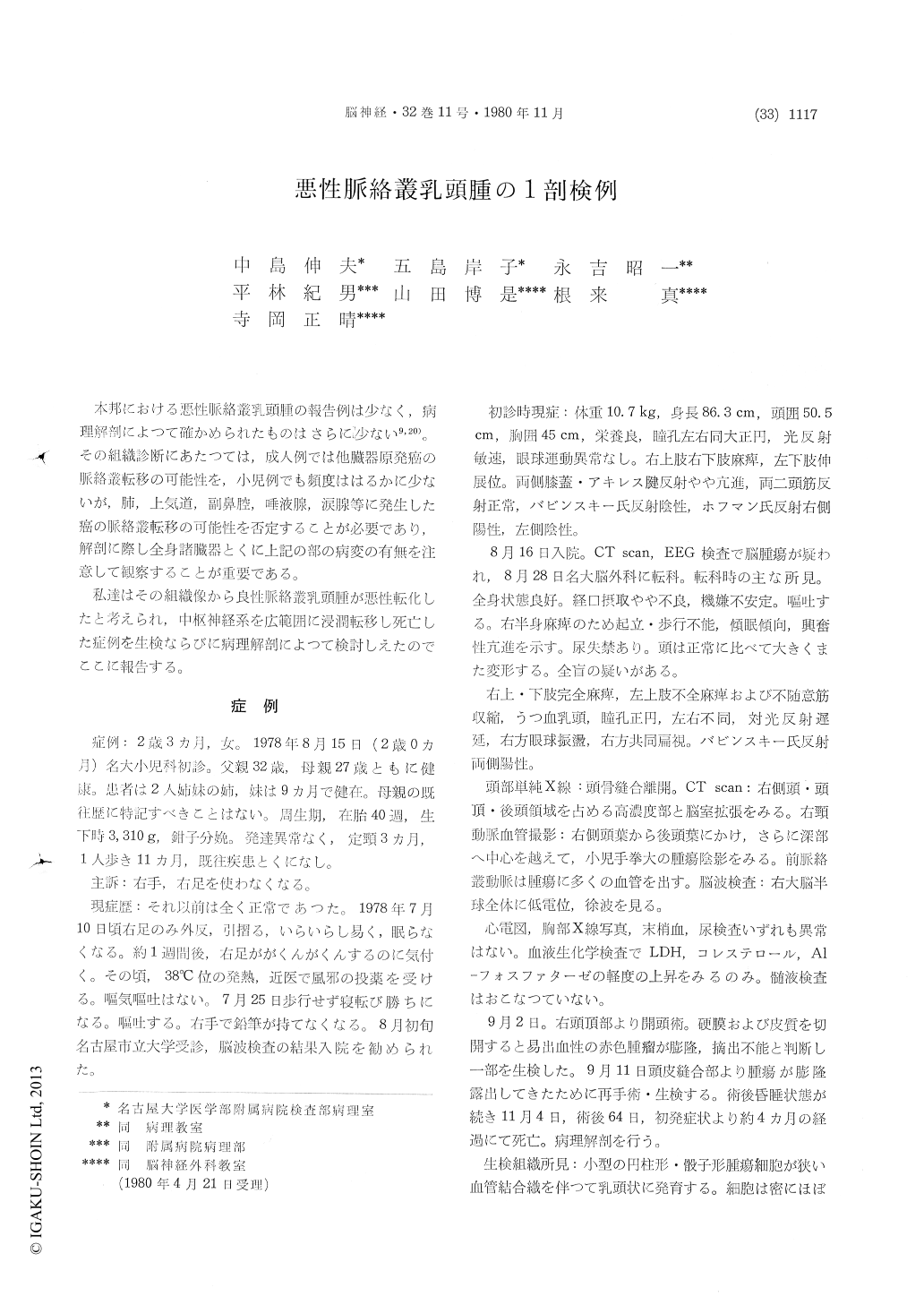
本邦における悪性脈絡叢乳頭腫の報告例は少なく,病理解剖によつて確かめられたものはさらに少ない9,20)。その組織診断にあたつては,成人例では他臓器原発癌の脈絡叢転移の可能性を,小児例でも頻度ははるかに少ないが,肺,上気道,副鼻腔,唾液腺,涙腺等に発生した癌の脈絡叢転移の可能性を否定することが必要であり,解剖に際し全身諸臓器とくに上記の部の病変の有無を注意して観察することが重要である。
私達はその組織像から良性脈絡叢乳頭腫が悪性転化したと考えられ,中枢神経系を広範囲に浸潤転移し死亡した症例を生検ならびに病理解剖によつて検討しえたのでここに報告する。
A rare autopsy case of malignant choroid plexus papilloma was reported. This 2 year and 3 months old girl had normal delivery history, and the deve-lopment and growth were not eventful. The patient admitted to Nagoya University Hospital because of motor disturbance of right extremities, in July, 1978. She vomited frequently and had the urinary incontinence.
Examinations by arteriograpy and CT Scanning revealed a tumor mass in the right cerebral hemisphere and basal nucleus. Findings from the hemogram, urinalysis, thoracic roentgenogram and electrocardiogram were all normal. Serum cholesterol, Al-phosphatase and lactic dehydrogenase activity were increased slightly. Cerebrospinal fluid was not examined.
Craniotomy was performed on September 2, whereby an elastic, red brown and easily hemorr-hagic tumor mass was found diffusely in the sub-cortical area of right parietal lobe, but the tumor was unresectable. Small biopsy specimens were removed for pathological examination of light and electron microscope. Eight days after the operation the tumor proliferated in the area of incison, and reoperated.
Light and electron microscopic examination of biopsies: The removed fragments of tumor consisted of a epithelial neoplasm of papillary structure with fine fibrovascular stroma. The epithelium was single layered, but mutilayering often occured. Moderate cellular atypism was found. There were many mitoses. Electronmicroscopically cillia and microvilli were rarely found. Malignant choroid plexus papilloma was suspected.
Autopsy findings: The relevant abnormal findings were confined to the central nervous system. Brain weighed 1,150gr. and showed marked defor-mity. The tumor originating in the right trigonum collaterale, invaded the surrounding brain sustanse, right basal nucleus and brain stem, and spreaded via cerebrospinal fluid widely. Pons and medulla oblongata were surronded and compressed by tumor. The tumor was grayish white in color, granular and spongy in consistence, and in the right cerebral hemisphere, large areas of yellowishwhite necrosis and bleeding were found. The third and forth ventricles were completely closed by pressure of the tumor. The frontal and inferior horns of right lateral ventricle were dilated respe-ctively.
Microscopical findings: In the right trigonum collaterale, where was supposed to be the primary lesion, the tumor was diagnosed benign choroid plexus papilloma histologically because of slight cellular and structural atypism and without mitoses. In the other invasive or metastatic lesion, the tumor showed features of malignant choroid plexus papilloma with marked cellular and structural atypism. Mitoses were found frequently.
This is the rare case to show that benign choroid plexus papilloma has the possibilities to change into malignant one.

Copyright © 1980, Igaku-Shoin Ltd. All rights reserved.


