Japanese
English
- 有料閲覧
- Abstract 文献概要
- 1ページ目 Look Inside
Ⅰ.はじめに
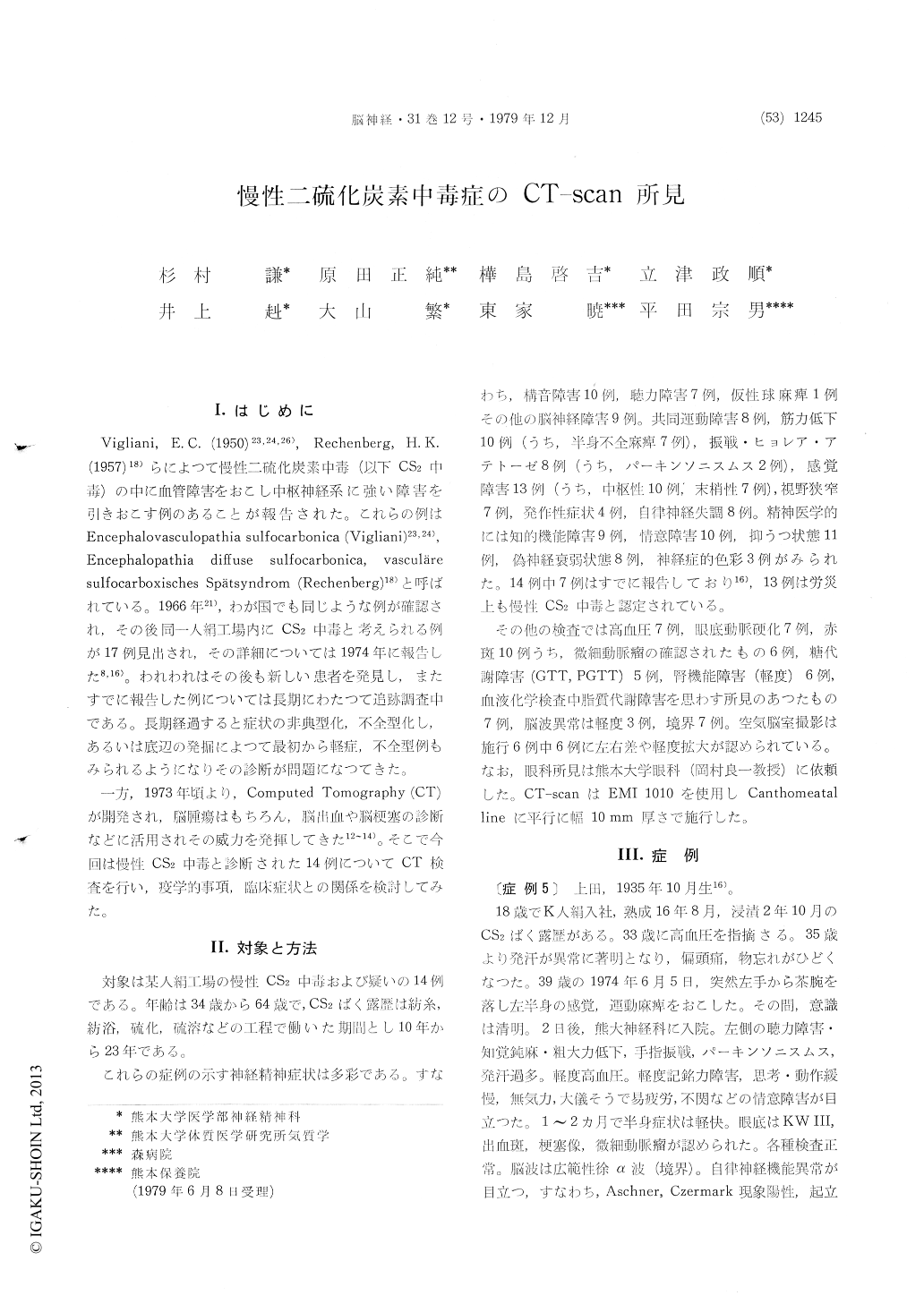
Vigliani,E.C.(1950)23,24,26),Rechenberg,H.K.(1957)18)らによつて慢性二硫化炭素中毒(以下CS2中毒)の中に血管障害をおこし中枢神経系に強い障害を引きおこす例のあることが報告された。これらの例はEncephalovasculopathia sulfocarbonica (Vigliani)23,24),Encephalopathia diffuse sulfocarbonica,vasculäresulfocarboxisches Spätsyndrom (Rechenberg)18)と呼ばれている。1966年21),わが国でも同じような例が確認され,その後同一人絹工場内にCS2中毒と考えられる例が17例見出され,その詳細については1974年に報告した8,16)。われわれはその後も新しい患者を発見し,またすでに報告した例については長期にわたつて追跡調査中である。長期経過すると症状の非典型化,不全型化し,あるいは底辺の発掘によつて最初から軽症,不全型例もみられるようになりその診断が問題になつてきた。
一方,1973年頃より,Computed Tomography (CT)が開発され,脳腫瘍はもちろん,脳出血や脳梗塞の診断などに活用されその威力を発揮してきた12〜14)。そこで今回は慢性CS2中毒と診断された14例についてCT検査を行い,疫学的事項,臨床症状との関係を検討してみた。
Vigliani, E. C.(1950)1), Rechenberg, H. K. (1957)2) et. al. have reported that in some cases of chronic carbon disulfide poisoning, vascular disorder causes severe damage to the central nervous system. In Japan similar cases found among rayon factory workers were reported in 1966, and in 1974 detailed symptoms of 17 cases were reported3). Meanwhile, diagnosis of cerebrovascular disease was advanced by the introduction of computerized tomography (CT).
Recently, CT examinations were made in 14 cases of chronic carbon disulfide (CS2) poisoning. The subjects were males ranging from 34 to 64 years of age. The range of duration of exposure to OS2 gas was from 10 years to 23 years. Clinical symp-toms were varied, with 9 cases of cranial nerve disturbance; 10 of dysarthria; 8 of incoordination ; 8 of muscular weakness ; 7 of hemiplegia ; and 13 of sensory disturbance. Among sensory disturbance,7 were of peripheral nerves and 10 of the CN. There were 7 cases of impaired hearing, 7 of con-striction of visual field, 4 of paroxysmal symptoms, 8 of vegitable symptoms, 9 of impairment of intel-ligence, 11 of personality change, 11 of depressive state, and 8 of neurasthenia.
Of the above, 13 cases were certified as chronic carbon disulfide poisoning in accordance with the Labor Accident Compensation Law. Recognized by clinical examination were 7 cases of hypertension, 7 of retinal arteriosclerosis, 10 of red dots on the retina, 6 of microaneurysma; 5 of saccharometabo-lism abnormality, 6 of abnormal renal function, 7 of abnormal lipoismetabolism, and 10 of EEG ab-normalities.
In observing CT, 10 cases of abnormality were found, eg., dilatation, 9 cases; dilatation of lateral ventricles, 6 cases; dilatation of the third ventricle, 1 case; dilatation of brain convulutions, 5 cases; dilatation of Sylvian fissure, 4 cases. Low density, which is assumed to be cerebral infarctions, was observed in 8 cases. Areas of low density were observed in caudale nucleus of basal ganglia in 3 cases; in the putamen 1 case; in the cortex 2 cases; and in the subcortex 4 cases.
Characteristic observations in CT of chronic CS2 poisoning were frequent images showing multiple infarction and brain atrophy. Of 4 cases without abnormal CT finding, 2 cases exhibited light clinica symptoms and in the other 2 cases various neuro-logical symptoms were observed. In 5 cases with severely abnormal CT findings clinical symtoms were also drastic, but in 1 case they were mild. That is, the degree of abnormality of CT-scan and severity of clinical symtoms are not correlated. Also, the location of abnormal CT findings does not coincide with the area of brain assumed to have been damaged on the basis of clinical symptoms. Differences in left and right and clinical symptoms coincide, and in 3 cases they were reversed. In five cases showing asymmetry in clinical symptoms, CT revealed no asymmetry. In each symptoms, 7 out of 9 cases were found to have serious disturb-ance of intelligence and strong atrophy of the frontal lobes. Of 4 cases with low density in the basal ganglia, extrapyramidal symptoms were absent in two cases; on the contraly, in 2 cases with parkinsonismus, low density in the basal ganglia was not detected.
In relation to other examination, CT coincides with clinical symptoms to a greater extent than EEG. Red dots on the retina and microaneurysma are more closely coorelated with clinical symtoms than is CT-scan. No relation between OS2 exposure duration and CT observation has been seen. In 8 out of 10 cases with stroke, abnormal CT was ob-served; and in 2 cases out of 4 without stroke, abnormal CT was obesrved. Also, the time lapse since stroke was 4 to 14 years in 10 cases. In two cases in which lapses were 9 years and 14 years respectively since stroke abnormal CT finding ab-normal was not observed. In 6 cases in which lapses were 4 to 7 years since stroke abnormal CT was observed. That is, generally speaking, cases with stroke but of relatively short history, had a tendency to show abnormal CT, but there were exceptions.
As an auxiliary method of diagnosing chronic CS2 poisoning, CT examination seems to be of future importance.
1) Vigliani, E. C.: Brit. J. Ind. Med. 11 : 235 (1954)
2) Rechenberg, H. K.: Arch. Gewerbepath. Ge-werbehyg. 15: 487 (1957)
3) Nakamura, K.& Harada, M.: Psychiat. Neu-rol. Jap. 76: 243 (1974)

Copyright © 1979, Igaku-Shoin Ltd. All rights reserved.


