Japanese
English
- 有料閲覧
- Abstract 文献概要
- 1ページ目 Look Inside
I.はじめに
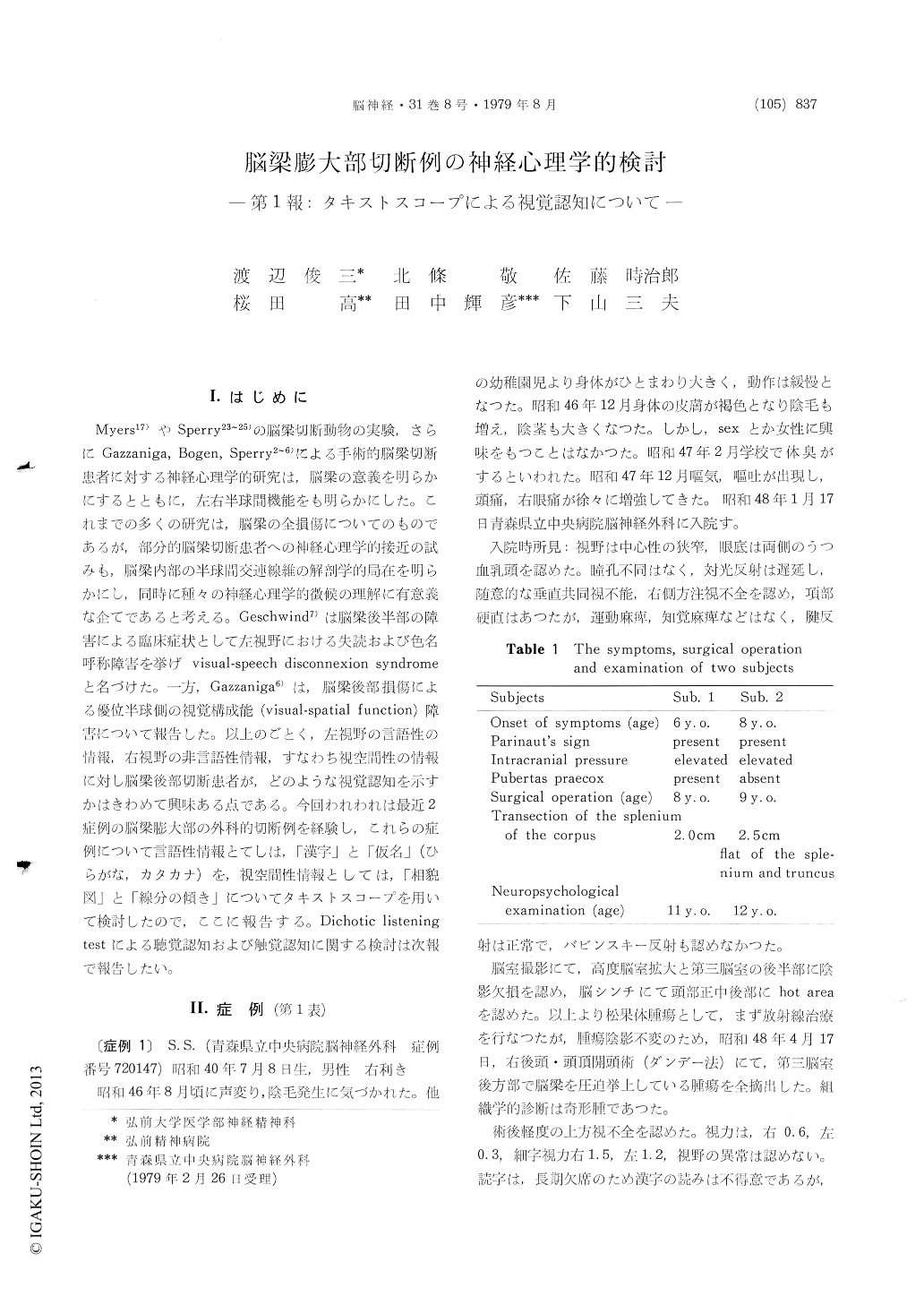
Myers17)やSperry23〜25)の脳梁切断動物の実験,さらにGazzaniga, Bogen, Sperry2〜6)による手術的脳梁切断患者に対する神経心理学的研究は,脳梁の意義を明らかにするとともに,左右半球間機能をも明らかにした。これまでの多くの研究は,脳梁の全損傷についてのものであるが,部分的脳梁切断患者への神経心理学的接近の試みも,脳梁内部の半球間交連線維の解剖学的局在を明らかにし,同時に種々の神経心理学的徴候の理解に有意義な企てであると考える。Geschwind7)は脳梁後半部の障害による臨床症状として左視野における失読および色名呼称障害を挙げvisual-speech disconnexion syndromeと名づけた。一方,Gazzaniga6)は,脳梁後部損傷による優位半球側の視覚構成能(visual-spatial function)障害について報告した。以上のごとく,左視野の言語性の情報,右視野の非言語性情報,すなわち視空間性の情報に対し脳梁後部切断患者が,どのような視覚認知を示すかはきわめて興味ある点である。今回われわれは最近2症例の脳梁膨大部の外科的切断例を経験し,これらの症例について言語性情報とてしは,「漢字」と「仮名」(ひらがな,カタカナ)を,視空間性情報としては,「相貌図」と「線分の傾き」についてタキストスコープを用いて検討したので,ここに報告する。Dichotic listeningtestによる聴覚認知および触覚認知に関する検討は次報で報告したい。
We made the neuropsychological visual studies of two right-handed subjects who had undergone the transection of the splenium of the corpus callosum for the pineal operations (section 2.0 cm and 2.5 cm). A binocular viewing, three-field Takei DP type of Tachistoscope for exposure duration from 80 to 120 msec was used to present the stimuli. The size of the stimuli were 1.5-2° (the visual angle), the presentation of the stimuli were presented 2°(the visual angle) to the right side and to the left side bilaterally. Stimulus material was composed with the various Japanese letters (Kana, Kanji), various faces (variation of the eyebrow form and the mouth form) and various slope of lines (interval of 10°).
Table 4 shows the results of two cases, the mean number of correct recognition in the right and left visual fields for the Kana, the Kanji, the face and the slope of the line tasks. It will be seen that performance of the subjects on the Kana and Kanji tasks showed a significant right field superiority, whereas on the face tasks there was left visual field superiority, but on the slope of the line there was no superiority between the right and the left visual fields. The task on the slope of the line was too difficult.
The Japanese orthography is unique in that two types of nonalphabetic symbols, Kana (phonetic symbols syllables) and Kanji (essentially nonphonetic logographic symbols representing lexical mor-phemes) are used in combinations. There was no definitive difference between the results of Kana and Kanji in our studies.
Our results show that the Kana and the Kanji (left hemisphere) and face (right hemisphere) are processed in fact differently in the cerebral hemi-sphere. The task on the slope of the line was too difficult for these subjects.

Copyright © 1979, Igaku-Shoin Ltd. All rights reserved.


