Japanese
English
- 有料閲覧
- Abstract 文献概要
- 1ページ目 Look Inside
I.はじめに
Westerberg3)(1966)以来,高血圧性脳出血例における脳血管写で,造影剤の脳血管外漏出像(extravasation)を認めたとの報告は多い4〜6,8)。しかし,これまでのところextravasationの出現時期,出血源,血腫形成機転についての報告が多く,extravasationの形状とその意義についての報告はわずかに高田8)にみるのみである。
著者らは外側型脳出血47例のうち,15例に血腫部位に一致したextravasationを認めたが,その形状を詳細に検討したところ,吉田,大根田らが原発性動脈破綻としているところの複数の線状体動脈に同時多発性に出血しているのを見い出した。この同時多発性出血を,脳血管写上にみとめたとの報告はいままでのところ文献上見当らない。
It is interesting to note the pathological findings that the rupture of multiple microaneurysms in the basal ganglia appears to be the cause of the hypertensive intracerebral hematoma. But there is few report that the carotid angiogram demon-strated multiple leakages of contrast medium of intracerebral hematoma in vivo than in Westberg's postmorten angiogram.
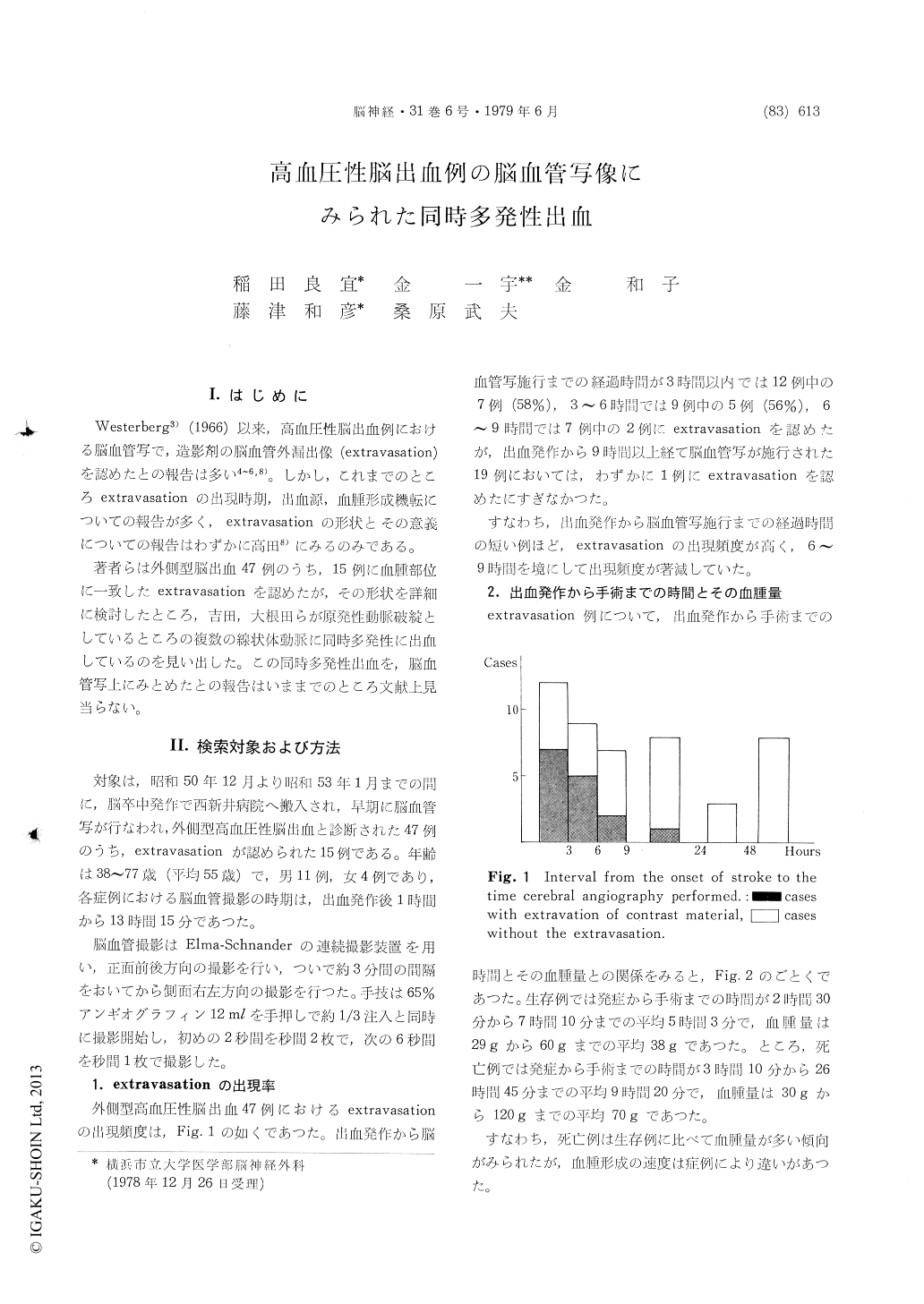
The authors experienced 15 cases of hypertensive intracerebral hematoma whose cerebral angiogram showed extravasations of contrast material. It was fiftyseven per cent of which angiogram within 6 hours after the stroke showed leakage of contrast medium.
However, there was remarkably decreased ap-pearence of the extravasation after 9 hours after the stroke.
Moreover, emphasis showed be placed upon thefacts that eleven cases of 15 cases showed more than two adjacent spotty extravasations of contrast medium and that six cases demonstrated the simultaneous multiple leakages of contrast material from more than two lentriculostriate arteries.
These angiographic findings has led us to the conclusion that there is intimate correlation between the large hematoma and multiple ruptures of the microaneurysms of lentriculostriate arteries.

Copyright © 1979, Igaku-Shoin Ltd. All rights reserved.


