Japanese
English
- 有料閲覧
- Abstract 文献概要
- 1ページ目 Look Inside
I.はじめに
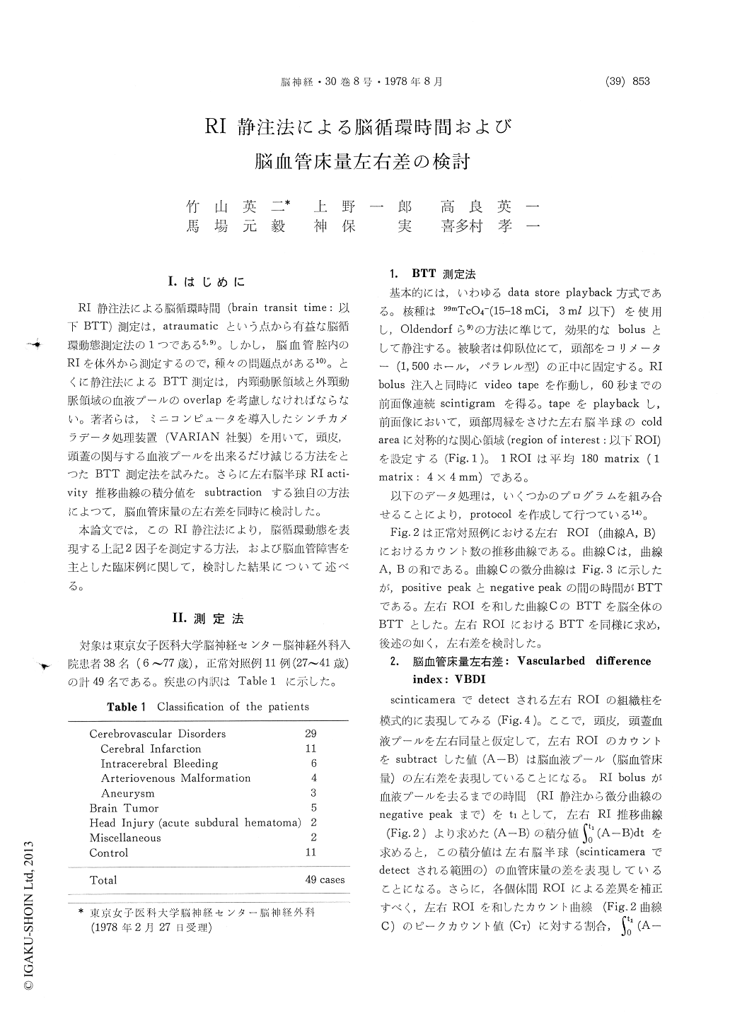
RI静注法による脳循環時間(brain transit time:以下BTT)測定は,atraumaticという点から有益な脳循環動態測定法の1つである5,9)。しかし,脳血管腔内のRIを体外から測定するので,種々の問題点がある10)。とくに静注法によるBTT測定は,内頸動脈領域と外頸動脈領域の血液プールのoverlapを考慮しなければならない。著者らは,ミニコンピュータを導入したシンチカメラデータ処理装置(VARIAN社製)を用いて,頭皮,頭蓋の関与する血液プールを出来るだけ減じる方法をとつたBTT測定法を試みた。さらに左右脳半球RI acti—vity推移曲線の積分値をsubtractionする独自の方法によつて,脳血管床量の左右差を同時に検討した。
本論文では,このRI静注法により,脳循環動態を表現する上記2因子を測定する方法,および脳血管障害を主とした臨床例に関して,検討した結果について述べる。
It will be accepted that by measuring radio-activity of the head after intravenous injection of RI some information could be afforded about vascular bed of the brain. However, external measurement method conventionally available at present includes inevitably some errors due to radioactivities in the extracerebral tissues, which make the analysis hard one in respect to extracting information about cerebral vascular bed selectively. To exclude these errors RI subtraction method could be a way of choice.
After intravenous injection of 99mTc-pertechnetate (15 mCi), radioactivities at the forehead were measured using gamma camera combined with a computer system. Two ROIs of about 30 cm2 were set symmetrically at the forehead and two count rate curves were obtained. Brain transit time (BTT) was calculated from first derivative of the initial count rate curve. As an index devoting difference between vascular bed of each hemisphere, the vascular bed difference index (VBDI) was introduced which was given by the following formula.
Where, A and B mean count rate curve obtained from non-affected and affected side of the head respectively. Thus integration of A (t) B (t) means difference from RI uptake between each cerebral hemisphere. Influence of radioactivities of the extracerebral tissues were theoretically extinguished by the subtraction. From technical reasons inte-grated interval was arbitrarily set between the time of RI injection (0) and negative peak of the first derivative curve (t1). As calibrating process the integrated value was divided by CT which is the highest count rate of total count rate curve synthesized from curve A and B.
BTT and VBDI were calculated in 49 subjects including 11 normal controls. In most of brain tumor, cerebral bleeding and severe head injury cases BTTs were prolonged in affected hemispheres. However in a half of cerebral infarction cases BTTs were rather shortened in affected sides.
In normal controls, absolute value of VBDI was|0.16±0.06 (SD)|. In cases of brain tumor, cere-bral hemorrhage and severe head injury, VBDI showed statistically significant deviation from normal range, taking plus value. This suggest, according to the definition of VBDI, vascular beds of affected hemisphere are abnormally decreasedcompared with those of non-affected side. On the other hand, in case of cerebral infarction, VBDI varies from case to case, making it difficult to get some conclusion from the present data.

Copyright © 1978, Igaku-Shoin Ltd. All rights reserved.


