Japanese
English
- 有料閲覧
- Abstract 文献概要
- 1ページ目 Look Inside
I.はじめに
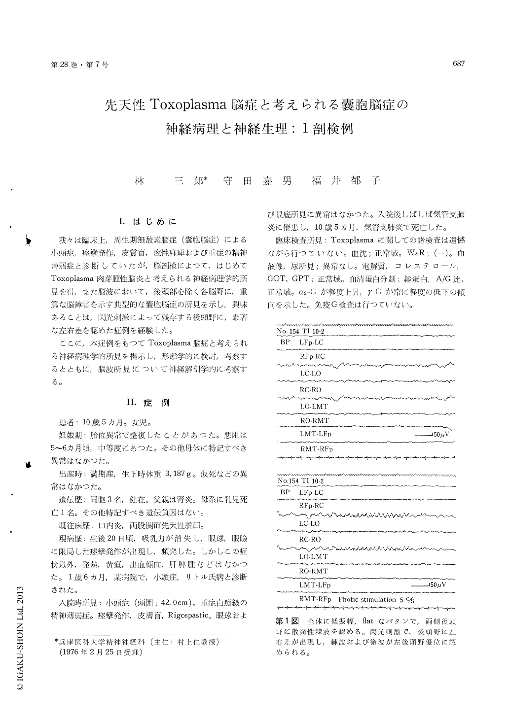
我々は臨床上,周生期無酸素脳症(嚢胞脳症)による小頭症,痙攣発作,皮質盲,痙性麻痺および重症の精神薄弱症と診断していたが,脳剖検によつて,はじめてToxoplasma肉芽腫性脳炎と考えられる神経病理学的所見を得,また脳波において,後頭部を除く各脳野に,重篤な脳障害を示す典型的な?胞脳症の所見を示し,興味あることは,閃光刺激によって残存する後頭野に,顕著な左右差を認めた症例を経験した。
ここに,本症例をもつてToxoplasma脳症と考えられる神経病理学的所見を提示し,形態学的に検討,考察するとともに,脳波所見について神経解剖学的に考察する。
An autopsy case of cystic encephalopathy with probable congenital toxoplasmic encephalopathy, together with interesting electroencephalogram was reported.
The patient was a girl aged 10 years 5 months. Family history was unremarkable and her siblings had been healthy. The pregnancy had been normal, and delivery was normal and at full term. Weight at birth was 3,187 g.
At 20 days after delivery she showed suddenly disability of sucking and focal ocular myoclonic seizure without other symptoms such as fever, vomiting, exanthema, chorioretinitis and hepatos-plenomegaly.
At the aged of 1, diagnosis of microcephaly and Little's disease was made. She suffered frequently from generalized epileptic seizure.
At the aged of 10, she was admitted to the hospital for severely handicapped. On admission, neurologi-cal and psychiatric examination revealed micro-cephaly, epileptic seizure, rigospastic state, cortical blindness and severe mental retardation, suspectedly due to multiple cystic encephalopathy of perinatal anoxic insult. Unfortunately, examination perti-nent to toxoplasma was not performed. The electroencephalogram showed low voltage and flat activity (almost complete electrical silence) in all areas, except on the occipital regions where single spike discharge. was seen. Intermittent photic stimulation elicited prominently asymmetrical change on the occipital regions which showed spikeand slow discharge on the left side.
She died of bronchopneumonia at the age of 10 years 5 months. Neuropathologically, the brain weighed 250 g. Extensive cystic cavities were seen in the entire cerebrum except the occipital regions, in the occipital lobe ulegyric cortical changes were noted in the medial parts of the right hemisphere, and cystic changes extended to some extent into the thalamus and periventricular zone. The lepto-meninges were hyperplastic but not infiltrated. Where the cortical and subcortical necrosis was marked, the neuronal architecture was completely destroyed and the proliferation of astrocytes and microglia, and glio-mesenchymal reaction forming scar, were noted. Furthermore, in these areas the blood vessel showed perivascular infiltrationof lymphocytes, plasma cells and round cells with an admixture of giant cells. Occasionally, endoarteritis obliterans and calcification of vessel walls were also seen in the necrotic tissue. The granulomatous inflammation was ubiquitously seen in the cerebrum, cerebellum, brain stem and spinal cord. Extensive pseudocalcification was also seen in these areas except the spinal cord. A large number of cysts with probable toxoplasma cyst were encountered in the neuronal parenchyma.
The clinico-neuropathological findings of cystic encephalopathy with probable congenital toxoplas-mic encephalopathy and clinico-anatomical correla-tion with electroencephalogram were briefly discussed.

Copyright © 1976, Igaku-Shoin Ltd. All rights reserved.


