Japanese
English
- 有料閲覧
- Abstract 文献概要
- 1ページ目 Look Inside
I.はじめに
トルコ鞍及びその近傍に発生する腫瘍の診断に際し,いくつかの補助診断法があるが,気脳撮影もその1つである。従来,どちらかと言えば血管撮影に関する所見の追求を主体とした研究が多く,気脳撮影による該部腫瘍の診断に関してはBakey2),Schaeffer3),Bull4),Twining5),Mahmoud6)等の報告があるが,いずれも鑑別診断に充分役立つとは言えない。
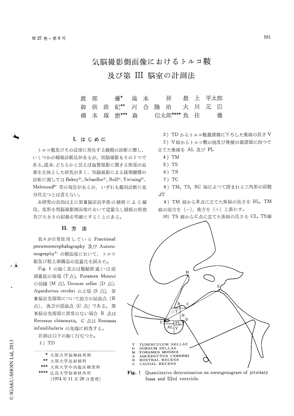
本研究の目的は主に第III脳室前半部の腫瘍による偏位,変形を気脳撮影側面像において定量化し腫瘍の形態及び大きさの記載を明確にすることにある。
1) The lateral projections of pneumoencephalo-gram were studied in normal cases, chromophobe adenomas, craniopharyngiomas, tuberculum sellae meningiomas and pinealomas in the chiasma region, with the reference points at the tuberculum sellae, the anterior wall of the Foramen of Monro, the entrance of aqueduct and the tip of the third ventricle.
2) In the 20 normal cases, the infundibular part of the third ventricle always stayed within the angle made by tuberculum sellae-Monro and tuberculum sellae-aqueduct line.
3) In chromophobe adenomas and acromegalies the size of the sella turcica was significantly larger than the controls.
4) In chromophobe adenomas with suprasellar extension, craniopharyngiomas and pinealomas in the chiasma region, it is confirmed that the anterior part of the third ventricle was indented superiorly and also anteriorly.
5) In tuberculum sellae meningiomas it is found that the anterior part of the third ventricle shows characteristic change. It is displaced posteriorly like pendulum.

Copyright © 1975, Igaku-Shoin Ltd. All rights reserved.


