Japanese
English
- 有料閲覧
- Abstract 文献概要
- 1ページ目 Look Inside
緒言
外傷性頭蓋内血腫の脳血管写像の読影に際して,最も困難を感ずるのは脳内血腫である。脳内血腫では,硬膜外血腫や硬膜下血腫のように無血管領野によつて確実な診断を下すことはできないので,脳血管の偏位のみによつて判断しなければならない。したがつて,Rousseauxら13)やLindgren8)は,脳内血腫の血管写像は他の頭蓋内占拠性病変,すなわち脳腫瘍などと同一であると記載しており,とくに頭部外傷に続発した場合には,限局性の脳浮腫との鑑別が困難となる。Petit-Dutaillis12)は,全般的な脳浮腫や限局性の脳挫傷を,脳内血腫と誤診した症例を報告している。
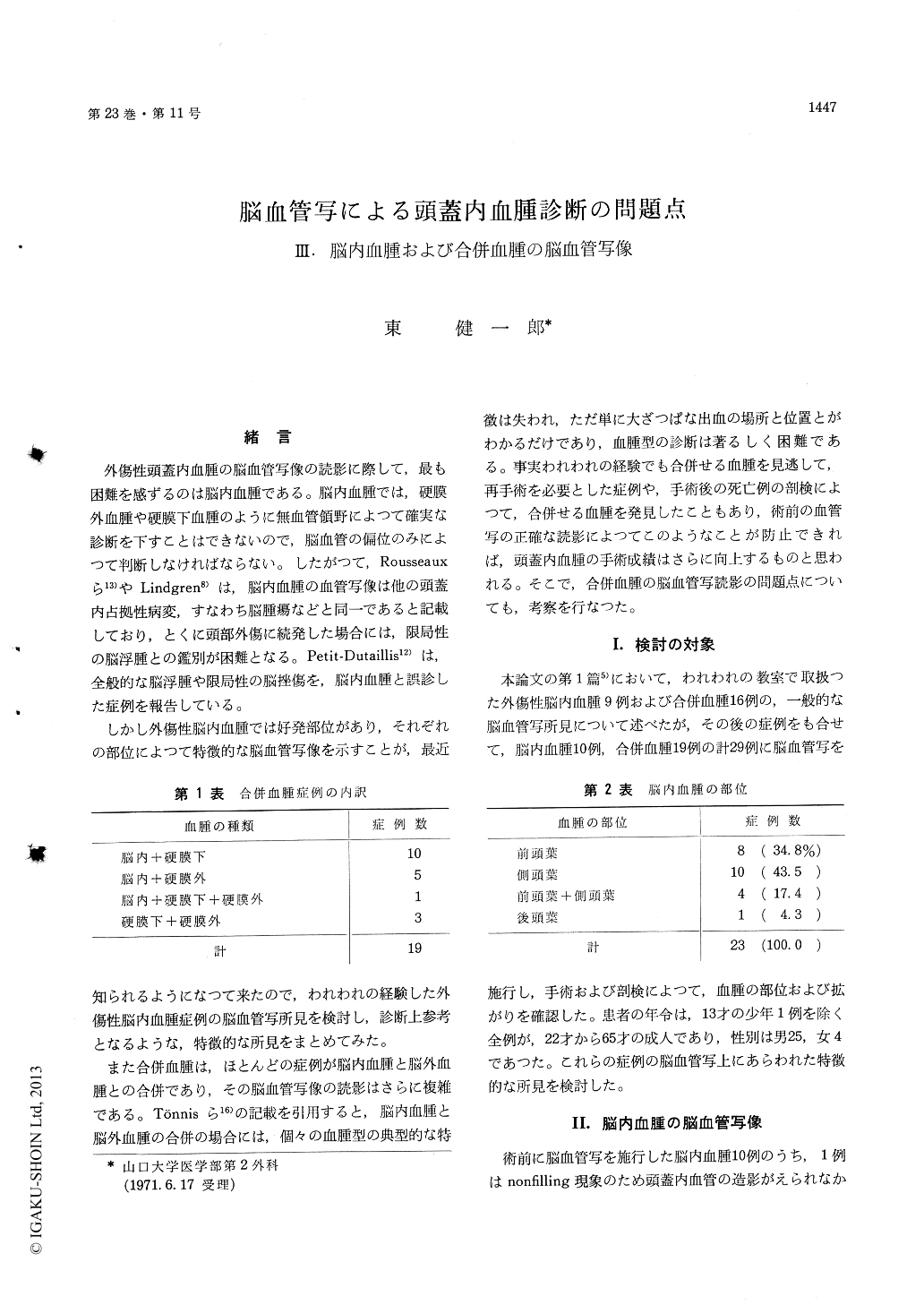
しかし外傷性脳内血腫では好発部位があり,それぞれの部位によつて特徴的な脳血管写像を示すことが,最近知られるようになつて来たので,われわれの経験した外傷性脳内血腫症例の脳血管写所見を検討し,診断上参考となるような,特徴的な所見をまとめてみた。
Twenty three cases of traumatic intracerebral hematoma were reviewed as to their angiographic findings. Among those cases, 14 had concomitant minor extracerebral hematomas, but their angio-graphic findings were thought to be mainly attri-butable to the intracerebral hematoma,
Intracerebral hematoma mainly develops in the frontal or temporal lobe. Hematoma in each site causes some characteristic features on the angiogram.
Unanimous findings in the frontal lobe hematoma are displacement of the anterior cerebral artery toward contralateral side and compressed shape of the carotid siphon in A-P view,and backwards dis-placement of the ascending branches of the middle cerebral artery and backwards compression of the sylvian triangle in lateral view.
Characteristic findings of the temporal lobe hematoma are upwards and medical displacement of the middle cerebral artery with straightening in A-P view, and forwards and upwards displace-ment of the middle cerebral artery with somewhat perpendicular course in its proximal portion in lateral view.
The courses of the Sylvian arteries on the lateral angiograms are different between two types of the hematomata ; the one mainly consists of blood clot and the other contains contused brain tissues as a principal ingredients. The main difference appears to be that the Sylvian arteries take straightened course in the former, while they preserve some tortuosity in the latter.
In general, however, angiographic features of intracerebral hematoma is similar to those of braintumor or other space-occupying lesion in the cor-responding site. And yet, differentiation between intra-and extracerebral hematoma is difficult in some cases.
Angiographic findings of multiple combined in-tracranial hematoma are diverse. Since the con-comitant hematoma is apt to be overlooked during surgery, careful scrutiny is necessary on the preo-perative angiograms.
Disproportion between the width of the midline shift of the anterior cerebral artery and that of avascular space or medial displacement of the skull helps the accurate diagnosis of combined hematoma on the same side.
Copyright © 1971, Igaku-Shoin Ltd. All rights reserved.