Japanese
English
- 有料閲覧
- Abstract 文献概要
- 1ページ目 Look Inside
緒言
頭部外傷患者の診断に際して最も重要なことは,頭蓋内血腫を早期に発見することである。臨床経過や神経学的所見から頭蓋内血腫の存在を予測できることもあり,超音波診断も有用ではあるが,最も正確に血腫の存在およびその部位まで診断できる方法は,脳血管写以外にはない。
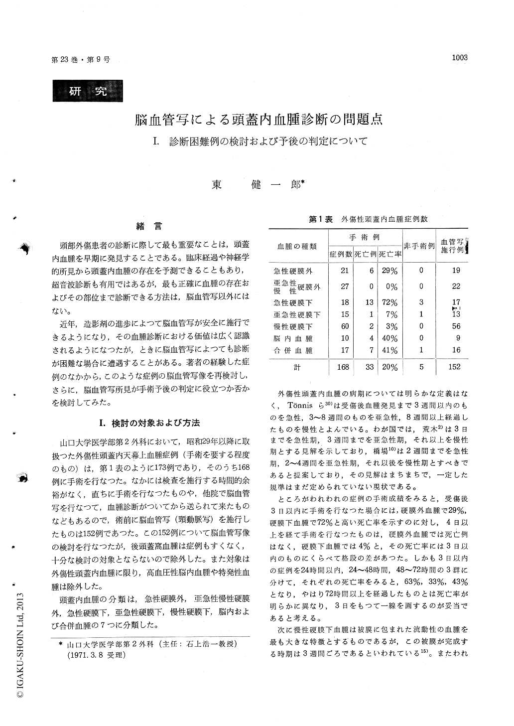
近年,造影剤の進歩によつて脳血管写が安全に施行できるようになり,その血腫診断における価値は広く認識されるようになつたが,ときに脳血管写によつても診断が困難な場合に遭遇することがある。著者の経験した症例のなかから,このような症例の脳血管写像を再検討し,さらに,脳血管写所見が手術予後の判定に役立つか否かを検討してみた。
Preoperative angiograms were studied on the 152 patients with traumatic intracranial hematoma. Dia-gnostic criteria for intracranial hematoma are made on angiograms according to following points ; (1) midline shift of the anterior cerebral artery or deep cerebral veins, (2) appearance of avascular space beneath the inner table of the skull, (3) abnormal course of the middle cerebral arterial group
Nonfilling phenomenon or delayed cerebral cir-culation is the most troublesome problem in angio-graphic diagnosis. These phenomena are thought to be due to an extremely high intracranial pres-sure and make visualization of above diagnostic signs impossible.
No opacification of the anterior cerebral artery is encountered in 10%, and no midline shift of thisartery, even if visualised, is found in 20% of the cases. Furthermore, avascular space is not visuali-ed in some cases on account of specific localization of hematomas or other reasons.
Such factors as mentioned above reduce the diagnostic value of cerebral angiogram. Possible causes and the polycy against above factors are discussed.
In 51 cases with acute traumatic intracranial hematoma, 23 alive and 28 dead, the rate of appear-ance of abnormal angiographic findings were studied. Consequently, following findings are considered to be useful for the estimation of prognosis, as indi-cating poor outcome ; nonfilling, delayed circulation, increase in the distance from the most lateral por-tion of the middle cerebral arterial stem to the inner table of the temporal bone in A-P arterio-gram, elevation of the middle cerebral artery, and downward displacement of the anterior choroidal artery. Downward displacement of the posterior cerebral artery and displacement of the venous angle are also useful signs indicating poor progno-sis.
If some of such findings suggesting poor pro-gnosis are visible on the preoperative angiogram, special care should be taken during surgery, such as an employment of extensive decompression.

Copyright © 1971, Igaku-Shoin Ltd. All rights reserved.


