Japanese
English
- 有料閲覧
- Abstract 文献概要
- 1ページ目 Look Inside
はじめに
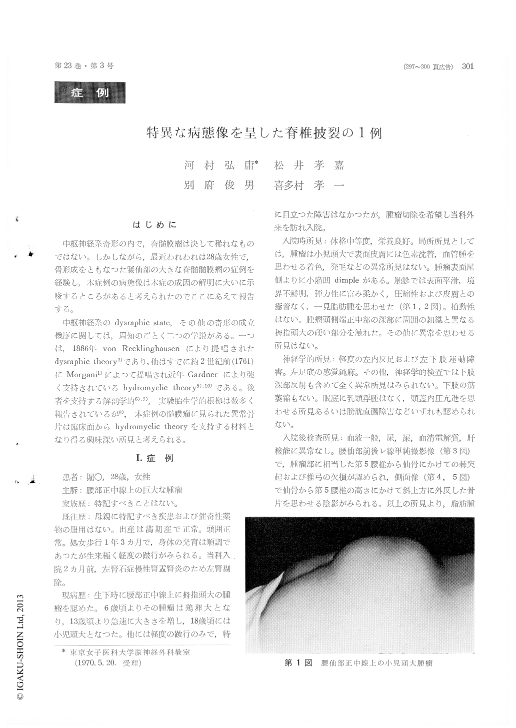
中枢神経系奇形の内で,脊髄膜瘤は決して稀れなものではない。しかしながら,最近われわれは28歳女性で,骨形成をともなつた腰仙部の大きな脊髄髄膜瘤の症例を経験し,本症例の病態像は本症の成因の解明に大いに示唆するところがあると考えられたのでここにあえて報告する。
中枢神経系のdysraphic state,その他の奇形の成立機序に関しては,周知のごとく二つの学説がある。一つは,1886年von Recklinghausenにより提唱されたdysraphic theory2)であり,他はすでに約2世紀前(1761)にMorgani1)によって提唱され近年Gardnerにより強く支持されているhydromyelic theory9),10)である。後者を支持する解剖学的6),7),実験胎生学的根拠は数多く報告されているが8),本症例の髄膜瘤に見られた異常骨片は臨床面からhydromyelic theoryを支持する材料となり得る興味深い所見と考えられる。
A case of lumbosacral atypical maningomyelocele associated with a bizarre bone flap was reported.
A 28-year-old female was admitted to our Depart-ment on Aug. 2, 1969, complaining of a huge mass in the lumbosacral region and slight disturbance of gait.
X-ray examinations of the lumbosacral spine revealed a spina bifida of L-5, S-1 and S-2 and an abnormal shadow which proved afterwards by operation to be a rare, abnormal bone.
At operation a large, flat, curved piece of bone was found in dome of the lumbosacral lipomatous tissue padding on the saccular meningomyelocele.
The bone flap (5cm × 8cm broad and 0. 7cm thick) had a punched out round hole with a smooth, thin edge in its central portion
Considering of the shape of the hole, it may be suggestive of one of the posterior sacral foramens and may be conceivably enough to explain that the bone may have arisen from the embryonal meso-dermal tissue developing lumbosacral bony struc-tures in normal case. In consequence, the bone evidently is different from the similar abnormal osteoid tissue of teratoma not infrequently associa-ted with spina bifida.
Concerning of the pathogenesis of the malfor-mations such as spina bifida, cranium bifidum and hydromyelia etc, hypotheses of von Recklinghau-sen's dysraphic theory and Morgagni's hydromye-lic theory have been held. If the findings of this case would be explained from dysraphic theory, it would have to be expected that the surrounding mesodermal structures, giving rise to -spinal proces-ses, laminas, muscles and connective tissue etc, resulted in genetic hypoplasia with failures of closure of the neural tube.
On the other hand, it seems to be reasonable that the bizarre bone having a characteristic round hole may have arisen from a heterotopic surround-ing mesodermal tissue migrated by overdistension of the neural tube from increased intraluminal pressure.
The authors, therefore, consider that these find-ings may have contributed not a little toward bringing Morgagni's hydromyelic theory (recently adovocated by Gardner) on genesis of spina bifida to support.
It is believed for us that coexistence of spina bifida and bone formation in this reported case may have been explained from a hydromyelia as sug-gested by Gardner.
The authors have briefly discussed the genetic cause of the spina bifida in considering of a coexistent strange bone of this case and reviewed reports of comparable cases.

Copyright © 1971, Igaku-Shoin Ltd. All rights reserved.


