Japanese
English
- 有料閲覧
- Abstract 文献概要
- 1ページ目 Look Inside
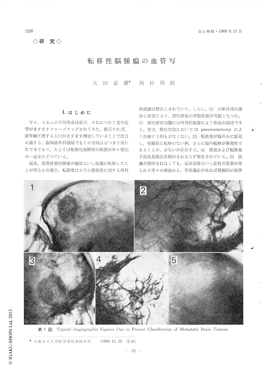
I.はじめに
年々,日本人の平均寿命は延び,それにつれて老年学がますますクローズアップされてきた。換言すれば,癌年齢に属する人口がますます増加していることで注目に値する。脳神経外科領域でもこの傾向ははつきり現われてきており,たとえば転移性脳腫瘍の頻度が年々増加の一途をたどつている。
従来,原発性悪性腫瘍が臓器ないし組織に転移したとが明らかな場合,転移巣はおろか原発巣に対する外科的侵襲は禁忌とされていた。しかし,(1)診断技術の進歩と啓蒙により,悪性腫瘍の早期発見が可能となつた。(2)悪性腫瘍初期には外科的処置により根治が期待できる。事実,肺癌初期においてはpneumonectomyにより治癒する例も少なくない。(3)転移巣が脳のみに限局し,他臓器に転移のない例,さらに脳内転移が単発性であることが,少ないが存在する。(4)原発および転移巣手術後長期生存例がまれならず報告されている。(5)治癒が期待されなくても,延命効果ないし症状の改善が得られる等々の理由から,手術適応があれば積極的に転移性脳腫瘍を手術するものが増えてきた。また,原発性脳腫瘍として手術し,術後組織診断で転移性腫瘍であることを発見し,原発巣をみいだすこともあれば,精力的諸検査にもかかわらず原発巣不明という例も多い。
Increased occurrence of the metastatic brain tumor has been closed up years by years and its propriety of surgical management has been discussed with incompatible data. This investigation has been undertaken in order to know whether or not metastatic brain tumors could be predicted preoperatively by angiographic characteristics of tumor stainings.
1) Forty-seven cases of metastatic brain tumors have been treated in out department and Kitano Hospital, Osaka. All of these cases have been diagnosed by a carotid angiography alone, so that metastasis in infratentorial space is excluded in our present analyses. Existance of space-occupying mass could be diagnosed angiographically in 38 out of 47 cases, and different patterns of tumor stains were noted in 17 out of 38 cases mentioned above (45%). Analysis of angiographic tumor stainings in 17 cases of metastatic brain tumors has been compared with those of glioblastoma multiforme and meningioma, because of angiographic similarity of their tumor stainings.
2) Pathologic stainings in a carotid angiography have been classified into 5 patterns (Table 1). The typical type in the metastatic brain tumor has been noticed as a diffusely spotty staining, while a roughly granular staining in glioblastoma multiforme and homogenously diffuse staining in meningioma (Table 3).
3) Circulatory velocity of contrast media in the tumor has been estimated in contrasting the densities of the tumor stainings in arterial, capillary and venous phases respectively. In case of glioblastoma multiforme the most dense tumor stain has been seen in arterial phase followed by a capillary phase. Tumor stain has already been disappeared in half cases of glioblastoma multiforme, while the most dense tumor stain in meningioma has been demonstrated in a capillary phase followed by a venous phase. In an arterial phase of meningioma tumor stain has not been seen yet in 8 out of 14 cases (57%) which means slow circulation in tumor mass.
The most dense tumor stain in case of the metastatic brain tumor has been demonstrated in capillary phase followed by an arterial and venous phases, which means intermediate circulatory velocity between glioblastoma multiforme and meningioma with closer similarity to cases of glioblastoma multiforme.
4) Above mentioned analyses have taught us that the typical characteristics of angiographic tumor stain in case of the metastatic brain tumor has been diffusely spotty in type and in relatively rapid circulation of contrast media. The arrangement of tumor stain, I (⧺) -I (⧻)-I (+) shown in Table 5 has been specifically seen in cases of the metastatic brain tumor, which, however, has been demonstrated in only 6 out of 17 cases (34%).
Copyright © 1969, Igaku-Shoin Ltd. All rights reserved.