Japanese
English
- 有料閲覧
- Abstract 文献概要
- 1ページ目 Look Inside
I.はじめに
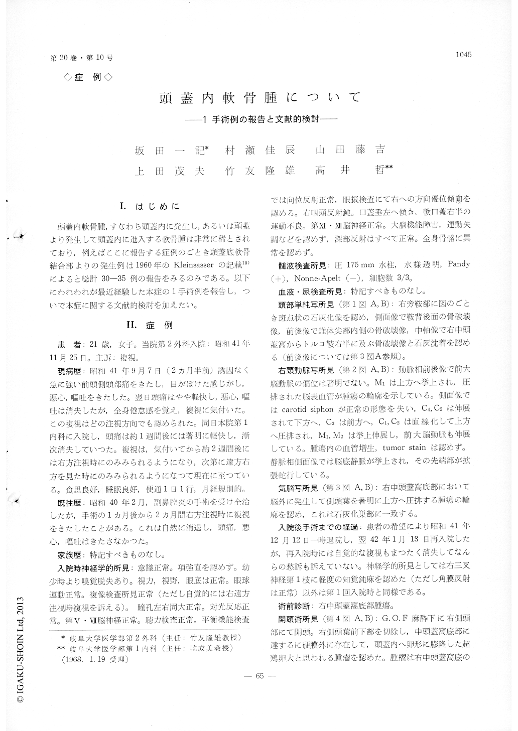
頭蓋内軟骨腫,すなわち頭蓋内に発生し,あるいは頭蓋より発生して頭蓋内に進入する軟骨腫は非常に稀とされており,例えばここに報告する症例のごとき頭蓋底軟骨結合部よりの発生例は1960年のKleinsasserの記載10)によると総計30-35例の報告をみるのみである。以下にわれわれが最近経験した本症の1手術例を報告し,ついで本症に関する文献的検討を加えたい。
Intracranial chondroma is one of the rare kinds of intracranial tumors and, according to the description by Kleinsasser in 196010), 30 to 35 cases in all of the chondroma arising from the region of the basal syn-chondroses had been reported by that time. Adding the cases reported thereafter, it seems that the total reported cases of this type of intracranial chondroma reach not more than about 45 cases at the present. We could not find any report thereon in Japanese literature. Recently we experienced a case of in-tracranial chondroma of the type arising from the basal synchondrosis region, which was subtotally re-moved operatively. The patient, a 21-year-old fe-male, suddenly complained of severe headache, nau-sea and vomitting 2 and a half months prior to admission to our clinic. These symptoms soon sub-sided, but she noticed diplopia, which was distinct on right gaze. By the time of the admission, how-ever, it almost subsided. Slight pareses of the right Vth, VIth (?), IXth and Xth cranial nerves were observed, but otherwise no neurological abnormlities were found. Spinal fluid pressure was within normal limit and papilledema was not found. Plain cranio-gram revealed an area of spotted calcification in the right parasellar region and there were bone destruc-tions in the right half of the sella turcica, posterior surface of the dorsum sellae and medial part of the temporal pyramid. Right carotid angiogram revealed a marked elongation and displacement of the in-tracranial internal carotid artery and elevation of the first portion of the right middle cerebral artery. Pneumoencephalogram showed the contour of a tumor in the right middle fossa. Right temporal craniotomy was performed and a middle fossa tumor exceeding a hen's egg size was removed subtotally within the capsule (dura mater). About 1 year after the operation the patient is healthy except for slight diplopia.
Number of cases of intracranial chondroma re-ported in the literature shows marked discrepancy between various reviewers. This seems to originate mainly from difference of definition and classification of this group of tumors. We consider that the clas-sification by Kleinsasser is most reasonable. Review of the literature on the tumor, especially of the type arising from the basal synchondrosis region was made and its incidence in intracranial tumors, age distribution, symptoms, diagnosis, roentgenologic fin-dings, histological findings, treatment and prognosis were discussed. Clinical symptoms may be very slight in spite of largeness of the tumor, as in the present case, and roentgenologic findings are be-lieved to he of utmost importance in its diagnosis. Operative removal is the only available treatment, but, since radical extirpation is almost always impos-sible, final prognosis is poor.

Copyright © 1968, Igaku-Shoin Ltd. All rights reserved.


