Japanese
English
- 有料閲覧
- Abstract 文献概要
- 1ページ目 Look Inside
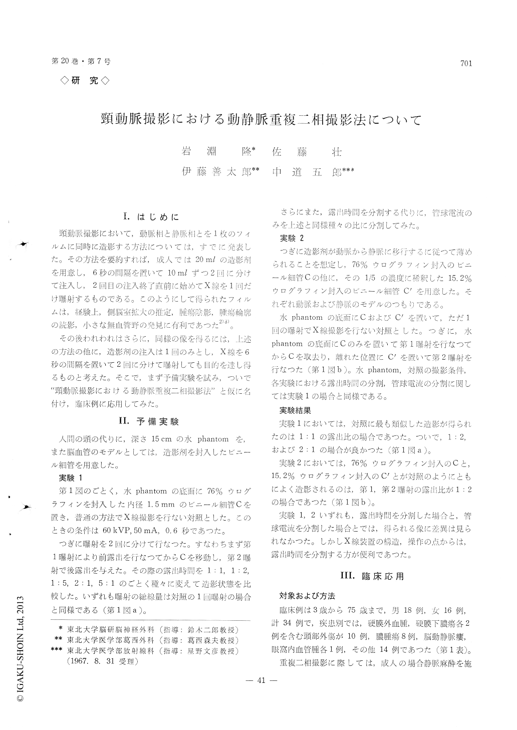
頸動脈撮影において,1回の造影剤の注入で,X線を2回に分けて曝射し,1枚のフィルムに動脈相と静脈相とを重ねて造影する方法を考え,予備実験の後34例の臨床例に試みた。
器具,手技,患者に対する侵襲は通常の頸動脈撮影の場合とまつたく同様で,動静脈各分枝をよく読影し得るフィルムが得られた。
動静脈重複二相撮影は,以前発表した同時二相撮影と同様に側脳室拡大の推定,腫瘍陰影,腫瘍輪廓の表現,小さな無血管野の発見,さらには無血管野の辺縁のより詳細な描出による硬膜上下の血腫と硬膜下膿瘍との鑑別などに有力であつた。
In carotid angiography, double exposure of the film following 2 injections of the contrast material at adequate interval was performed in an effort to obtain both arterial and venous phases on a single film. Following preliminary experiment 34 clinical cases were subjected to the study. Instruments, technique and influence on patients were quite similar to those for ordinary carotid angiography, thus fascilitating diagnostic interpretation of each tribu-tary of arterial and venous phases. The two phase carotid angiopolygraphy was of great value along with the previously reported simultaneous two phase carotid angiography for determination of tumor stain, outline of the tumor, presumption of dilatation of the lateral ventricles and differentiation of extra and/or subdural hematoma from subdural abscess by demonstrating of the margin of an avascular area.

Copyright © 1968, Igaku-Shoin Ltd. All rights reserved.


