Japanese
English
- 有料閲覧
- Abstract 文献概要
- 1ページ目 Look Inside
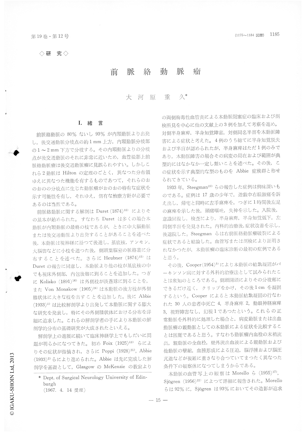
I.緒言
前脈絡動脈の80%ないし93%が内頸動脈より出発し,後交通動脈分岐点の約1mm上方,内頸動脈分岐部の1〜2mm下方で分岐する。その内頸動脈よりの分岐点が後交通動脈のそれに非常に近いため,血管撮影上前脈絡動脈瘤は後交通動脈瘤に見誤られやすい。しかしこれら2動脈はHiltonの定理のごとく,異なつた分布領ゆえに異なつた機能を有するものであつて,それらのおのおのの分岐点に生じた動脈瘤がおのおの特有な症状を示す可能性を有し,それゆえ,個有な治療方針が必要であるのは当然である。
前脈絡動脈に関する解剖はDuret (1874)10)によりその基本が始められた。すなわちDuretは多くの場合本動脈が内頸動脈の最終の枝であるが,ときに中大脳動脈または後交通動脈より出発することがあることを述べた後,本動脈は視神経に沿つて後進し,基底核,アンモン,大脳脚などに小枝を送つた後,側頭葉脳室の脈絡叢に分布することを述べた。さらにHeubner (1874)15)はDuretの報告に同意し,本動脈より他の枝が基底核の中でも視床外側部,内包後脚に到ることを追加した。つぎにKolisko (1891)16)は外側枝が淡蒼球に到ることを,またVon Monakow (1905)24)は本動脈の後方枝が外側膝状体に大きな渋を出すことを追加した。後にAbbie(1933)1)は比較解剖学より出発して本動脈に関する膨大な研究を発表し,特にその外側膝状体における分布を詳細に追求した。これらの解剖学者の手により本動脈の解剖学的分命の基礎研究が大成されたといえる。
Aneurysm of the anterior choroidal artery is relatively uncommon and ranges from 1 to 2% sta-tistical incidences reported by rather few authors. At first in this paper the historical development of anatomical and pathological knowledge on this artery is reviewed.
Four cases of ruptured aneurysm of the anterior choroidal artery are discribed in this paper. Two out of four cases showed Abbie's syndrome, which consists of contralateral motor, sensory impairments and hemianopsia. This syndrome is to be explained on the basis of its pathological changes at the pos-terior limb of the internal capsule, crus cerebri, lateral geniculate body, optic tract and radiation normally supplied by numerous branches from the anterior choroidal artery. To seek this syndrome and confirm on it could be very helpful for diagnosis of this aneurysm, especially so in case relevant an-giographical studies turn out inconclusive for its diagnosis. On these four cases impairment of ex-ternal ocular movements was not found, which is contrarily quite common for aneurysm of the pos-terior communicating artery. Various types of visual field impairments are also discribed to be very helpful for localization of pathology caused by rupture of this aneurysm and therefore for its diagnosis.
The relevant phylogenetical, anatomical and ra-diological details on the anterior choroidal artery are discribed.
On antero-posterior view of angiography this a-neurysm tends to reveal itself as a medially directed protrusion from C-1 portion of the internal carotid artery, and aneurysm of the posterior communicat-ing artery, on the contrary, tends to protrude later-ally on angiography. On lateral view it is cruciallyimportant for diagnosis of this aneurysm to identify the origin of the posterior communicating artery.The aneurysm is occasionally so large that it might be conceivably overshadow on both origins of the anterior choroidal and posterior communicating arter-ies. In such a case oblique view might be helpful to identify one origin from another for diagnosis there.
On its treatment careful planning should be made on the basis of results from various kinds of cross circulation tests. When satisfactory cross circulation is proved to exist ligation of the carotid artery would be method of choice. When a good cross circula-tion is unfortunately denied direct approach should be planned to prevent postoperative complication as soon as patient's condition reaches sufficiently enough to tolerate the procedure. On the contrary to an aneurysm of the posterior communicating artery which is essentially bridge artery without much direct blood supply to vital brain structures and allowes to be ligated usually without much untoward effects, the parent arteay of this aneurysm, anterior choroidal artery should not be ligated because of its direct blood supply to the vital brain structures in spite of its strong anastomosis with mainly the posterior choroidal artery. It should be wrapped or coated so as to keep its blood stream in normal fashion.
In most of up to date aeports this aneurysm has not been clearly identified in its diagnosis, symp-tomatology or teatment. It is emphasized in this paper that this aneurysm should be clearly identified and dealed with differently from one at the posterior communicating artery junction.

Copyright © 1967, Igaku-Shoin Ltd. All rights reserved.


