Japanese
English
- 有料閲覧
- Abstract 文献概要
- 1ページ目 Look Inside
I.はじめに
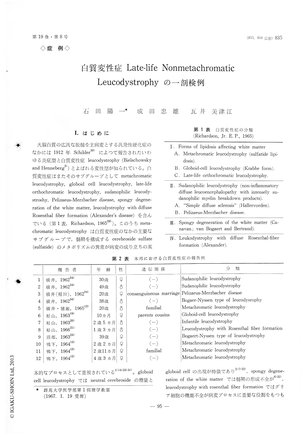
大脳白質の広汎な脱髄を主病変とする汎発性硬化症のなかには1912年Schilder30)によつて報告されたいわゆる炎症型と白質変性症leucodystrophy (Bielschowskyand Henneberg3))とよばれる変性型が知られている。白質変性症はまたそのサブグループとしてmetachromatic leucodystrophy, globoid cell leucodystrophy, late-life orthochromatic leucodystrophy,sudanophilic leucody—stroshy, Pellzaeus-Merzbacher disease, spongy degene—ration of the white matter, leucodystrophy with diffuse Rosenthal fiber formation (Alexander's disease)を含んでいる(第1表,Richardson,196529))。このうちmeta—chromatic leucodystrophyは白質変性症のなかの主要なサブグループで,髄鞘を構成するcerebroside sulfate(sulfatide)のメタボリズムの異常が病変の成り立ちの基本的なプロセスとして重視されている1)14)23)31)。globoid cell leucodystrophyではneutral cerebrosideの増量とgloboid cellの出現が特徴であり2)7)22),spongy degene—ration of the white matterでは髄鞘の形成不全が6)32),leucodystrophy with rosenthal fiber formationではグリア細胞の機能不全が病変プロセスに重要な役割をもつものと考えられている29)。orthochromatic leucodystrophy (sudanophile leucodystrophy)では脱髄巣内にみられる清掃細胞にとりこまれた解体物質はズダン好性で,髄鞘のカタボリズムに異常を示す所見に乏しく,症例のなかには血管炎所見の有無を別とするとSchilder型脳炎と区別困難な場合もある。この型のleucodystrophyにもeinfache orthochromatische Leukodystrophie (Pfeiffer25)) (sudanophile leucodystrophy;Greenfield and Norman,9) einfache degenerative Leukodystrophie;Hallervordenlo)),Pelizaeus-Merzbacher disease,8)18)late-life orthochromaticleucodystrophy29)などのサブグループがある。本邦でも1960年以来,白質変性症の報告例は12例を数え,meta—chromatic leucodystrophy (鴨下,浦野15)),globoid cell leucodystrophy (松山,入20)) leucodystrophy with diffuse rosenthal fiber formation (松山,入21))の諸型が記載されているが,また横井の3例34),Bogaert-Nyssen typeとして記載された沼部,岸24)例のように晩発性のortho—chromatic leucodystrophyの記載が多い。ここに報告する例も晩発性の白質変性症の1例で,病理組織学的,組織化学的,生化学的検索を行なつたものである。
Histopathologic investigation of a case of leucody-strophy is presented in a case who died at the age of 57 years. The illness developed during adult life and had a slowly progressive prolonged clinical course of approximately 7 years. Disturbaces in articulation, muscular rigidity and mental deteriolation were the dominant clinical features. Autopsy revealed an widespread bilateral greyish gelatinous appearance of the cerebral white matter with sparing of the narrow subcortical layer of U fibers. Sections stained for myelin showed diffuse bilateral pallor of the cerebral hemispheres extending througout the centrum ovale and corpus callosum. Holzer stain showed diffuse dense gliosis of the demyelinated areas. A conside-rable loss of axis cylinders was also seen with the formation of many spherical end-bulbes. Perivascular inflammatory cell reaction was minimal or allmost absent. Microglial phagocytes were not abundant and found only scattered throughout the demyelinated area. They contained sudanophil lipid and occasio-nally nonmetachromatic feebly sudanophil materials or light brown pigment staining positively for iron. Histopathological and histochemical examinations sug-gest that this case belongs to the category of suda-nophile leucodystrophy, particularly of late-life ortho chromatic leucodystrophy. Chemical analysis showed decrease in amount of phospholipid, glycolipid, and cholesterol with an increase in amount of esterified cholesterol. No evidence of defective myelin cata-bolism was demonstrated in the demyelinated tissue of this case.

Copyright © 1967, Igaku-Shoin Ltd. All rights reserved.


