Japanese
English
- 有料閲覧
- Abstract 文献概要
- 1ページ目 Look Inside
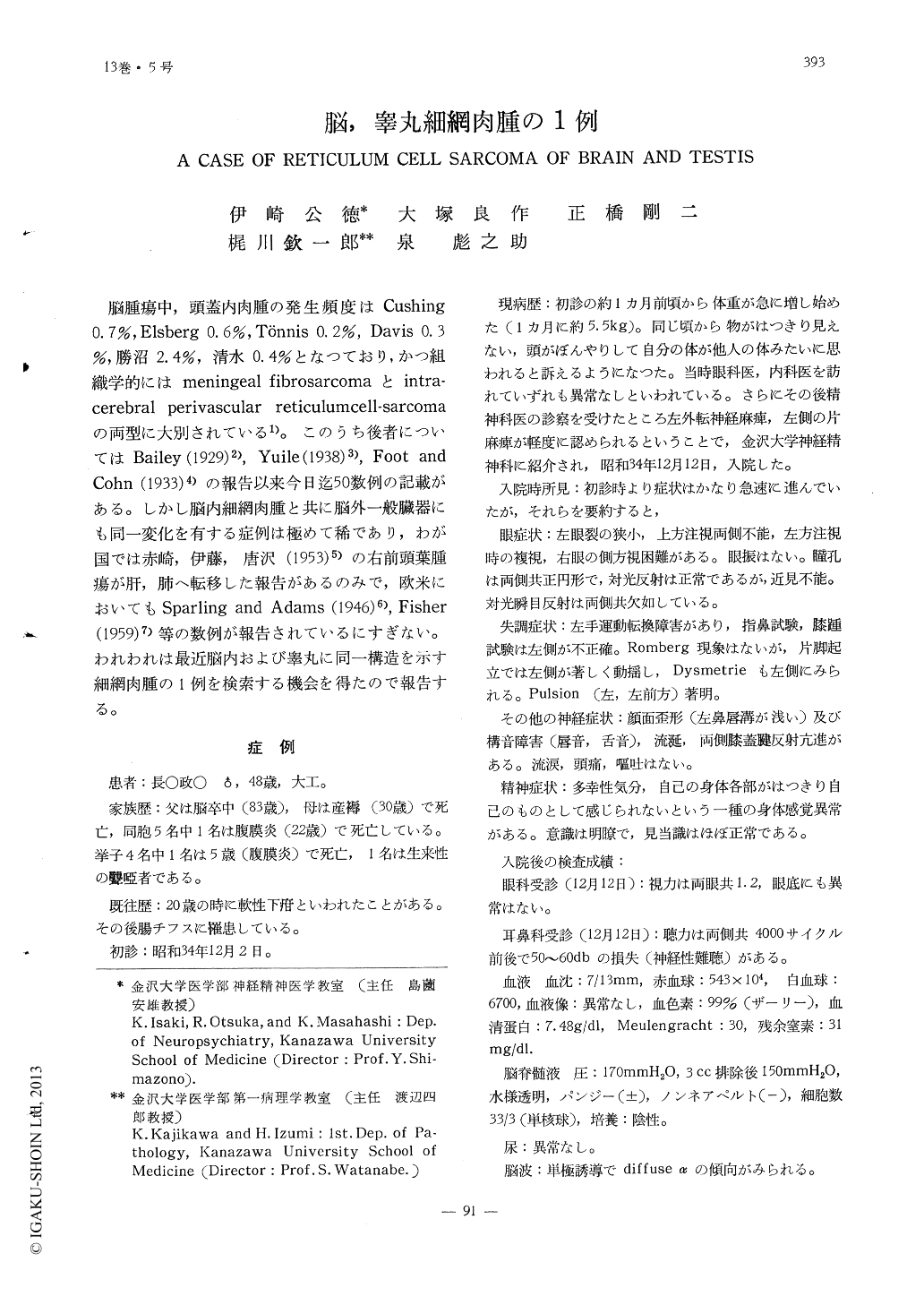
体重増加,眼球運動障害,運動失調をもつて発病し,剖検の結果脳及び左睾丸に細網肉腫を認めた1例(48歳の男子)を報告した。
脳腫瘍は肉眼的には視束交叉に中心をもち,前方は右被殻前極,上方には右側視床,被殻を上,外方に破壊性に圧迫し,後方は大脳脚に沿つて脳橋右半側,第四脳室周辺まで連続した発育をし,さらに顕微鏡的には左視床,小脳,延髄の諸核及び脳底軟膜にも広く浸潤していた。
左睾丸は外見上変りないが,割面では淡紅灰白色を呈し,正常構造が認められない。
腫瘍細胞は泡状核の長円形又は不整円形の短突起を有する細胞や,クロマチンに富む円形細胞よりなり,かなりの核分裂を示しているが,グリア系の細胞ではなかつた。腫瘍内の線維形成はかなりみられ貧喰能が著しく,特に血管周囲性の増殖が顕著である。
このような所見からわれわれは本症を細網肉腫の1例とみなし,本腫瘍とHortegaグリア腫瘍との関連について考察した。
A case of reticulum cell sarcoma of brain, with identical histopathologic focus in one testis, was reported.
The patient, a 48 years old carpenter, had increase in weight about 2kg for a month, and complained disturbance of vision and dy-sbasia. Thereafter, ataxia, disturbance of eye-movement, and mania-like sympotomes oc-cured, but no headache or vomiting. Diag-nosis of the tumor in the midbrain was sug-gested, however, there was no abnormal fin-dings in the X-ray examination, spinal fluid, and other laboratory studies.
Necropsy revealed a soft mass measuring 3×3 cm at the chiasma fasciculorum opticum. By coronal sections of the brain, macrcsco-pically it disclosed homogeneous, gray-white tumor tissure, extending anteriorly from the rostal portion of the right caudate nucleus and posteriorly to the pons throught the ri-ght midbrain, and with microscopic examina-tion tumor cells infiltrated into the left thal-mus and the adjucent of the fourth ventricle.

Copyright © 1961, Igaku-Shoin Ltd. All rights reserved.


