Japanese
English
- 有料閲覧
- Abstract 文献概要
- 1ページ目 Look Inside
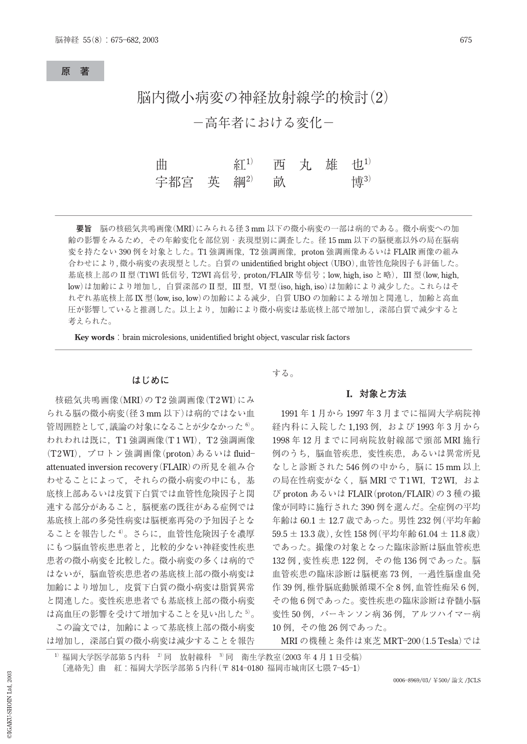
要旨 脳の核磁気共鳴画像(MRI)にみられる径3mm以下の微小病変の一部は病的である。微小病変への加齢の影響をみるため,その年齢変化を部位別・表現型別に調査した。径15mm以下の脳梗塞以外の局在脳病変を持たない390例を対象とした。T1強調画像,T2強調画像,proton強調画像あるいはFLAIR画像の組み合わせにより,微小病変の表現型とした。白質のunidentified bright object (UBO),血管性危険因子も評価した。基底核上部のII型(T1WI低信号, T2WI高信号, proton/FLAIR等信号; low, high, isoと略),III型(low, high, low)は加齢により増加し,白質深部のII型,III型,VI型(iso, high, iso)は加齢により減少した。これらはそれぞれ基底核上部IX型(low, iso, low)の加齢による減少,白質UBOの加齢による増加と関連し,加齢と高血圧が影響していると推測した。以上より,加齢により微小病変は基底核上部で増加し,深部白質で減少すると考えられた。
Background : Some of the cerebral microlesions less than 3mm in diameter observed on magnetic resonance imaging(MRI)are considered to represent pathological processes. The present study investigated changes due to aging in microlesions according to anatomical regions and phenotypes. Patients : A total of 390 cases without localized lesions other than lacune less than 15mm in diameter were studied. Method : Microlesion type on MRI was categorized into hypo-, iso-, and hyper-intensities on T1-weighted(T1WI), T 2-weighted(T2WI), and proton-weighted or fluid-attenuated inversion recovery(proton/FLAIR)images. Correlations between unidentified bright objects(UBO)in white matter and vascular risk factors were analyzed using logistic regression analysis. Results : Microlesions of the upper basal ganglia showing low intensity on T1WI, high intensity on T2WI and iso intensity on proton/FLAIR, and showing low intensity on T1WI, high intensity on T2WI and low intensity on proton/FLAIR increased with age, whereas those showing low intensity on T1WI, iso intensity on T2WI, and low intensity on proton/FLAIR of the upper basal ganglia decreased. In subcortical white matter, microlesions of the first two types : and those showing iso intensity on T1WI, high intensity on T2WI and iso intensity on proton/FLAIR decreased with age. Conversely, UBO increased with age, and significantly correlated with hypertension. Conclusion : Although microlesions in the upper basal ganglia increase with age, those in the subcortical white matter decrease with age. These observations suggest pathological changes surrounding small arteries with aging in the brain.

Copyright © 2003, Igaku-Shoin Ltd. All rights reserved.


