Japanese
English
- 有料閲覧
- Abstract 文献概要
- 1ページ目 Look Inside
- 参考文献 Reference
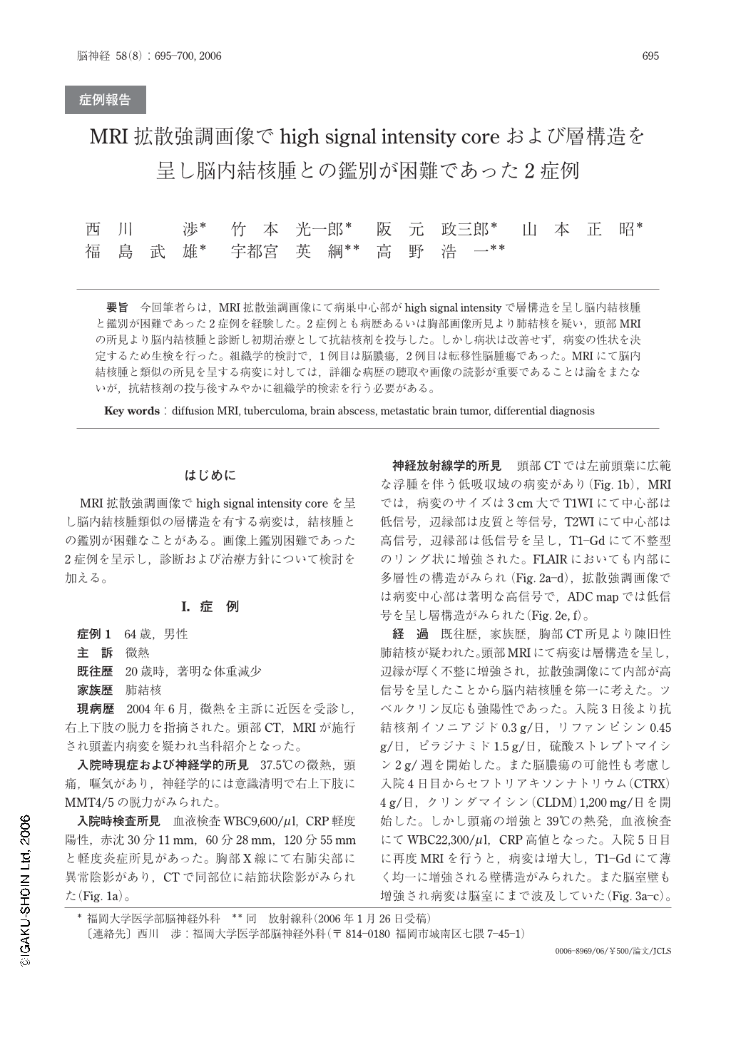
要旨 今回筆者らは,MRI拡散強調画像にて病巣中心部がhigh signal intensityで層構造を呈し脳内結核腫と鑑別が困難であった2症例を経験した。2症例とも病歴あるいは胸部画像所見より肺結核を疑い,頭部MRIの所見より脳内結核腫と診断し初期治療として抗結核剤を投与した。しかし病状は改善せず,病変の性状を決定するため生検を行った。組織学的検討で,1例目は脳膿瘍,2例目は転移性脳腫瘍であった。MRIにて脳内結核腫と類似の所見を呈する病変に対しては,詳細な病歴の聴取や画像の読影が重要であることは論をまたないが,抗結核剤の投与後すみやかに組織学的検索を行う必要がある。
Two cases with a high signal intensity core with laminar structure based on diffusion weighed-images (DWI) mimicking intracerebral tuberculoma were herein reported. Both patients were suspected to have pulmonary tuberculosis in their past history and/or based on chest X-ray findings. DWI demonstrated a high signal intensity core with laminar structure, thus suggesting the presence of an intracerebral tuberculoma. As initial therapy, both patients received anti-tuberculous drugs, however, their symptoms did not improve, and the lesions were observed to have increased in MRI. As a result, a biopsy was carried out to clarify the nature of the disease. A pathological examination showed the former to be a brain abscess while the latter was metastatic carcinoma. In such cases mimicking intracerbral tuberculoma based on DWI, anti-tuberculous drugs are first treatment of choice, however, a biopsy should be performed as early as possible if the medication dose not show a significant response.

Copyright © 2006, Igaku-Shoin Ltd. All rights reserved.


