Japanese
English
- 有料閲覧
- Abstract 文献概要
- 1ページ目 Look Inside
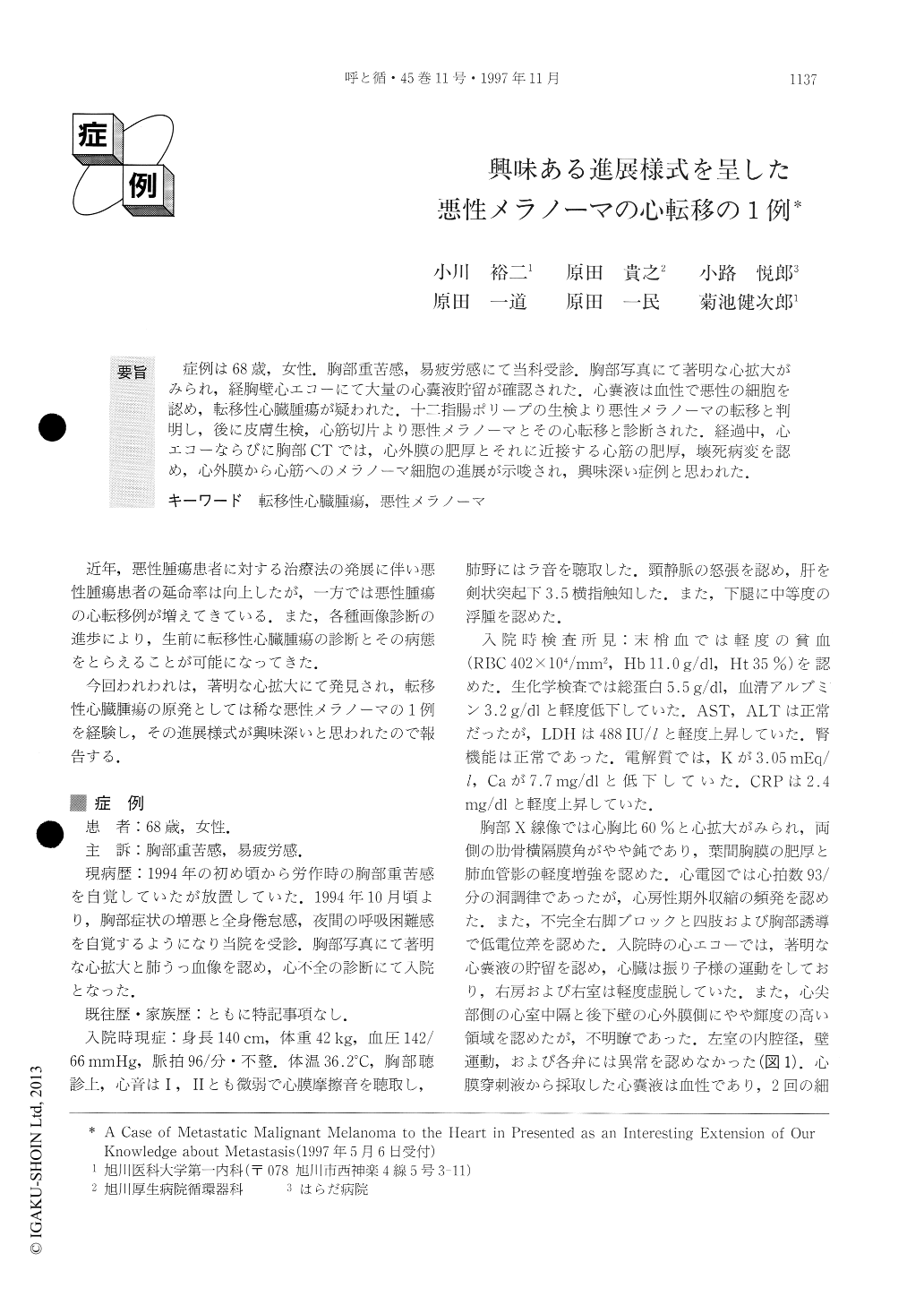
症例は68歳,女性.胸部重苦感,易疲労感にて当科受診.胸部写真にて著明な心拡大がみられ,経胸壁心エコーにて大量の心嚢液貯留が確認された.心嚢液は血性で悪性の細胞を認め,転移性心臓腫瘍が疑われた.十二指腸ポリープの生検より悪性メラノーマの転移と判明し,後に皮膚生検,心筋切片より悪性メラノーマとその心転移と診断された.経過中,心エコーならびに胸部CTでは,心外膜の肥厚とそれに近接する心筋の肥厚,壊死病変を認め,心外膜から心筋へのメラノーマ細胞の進展が示唆され,興味深い症例と思われた.
A 68-year-old woman had experienced chest oppres-sion and fatigue. A chest X-ray film showed cardiac enlargement (CTR 60%). Transthoracic echocardiogra-phy (TTE) revealed a large pericardial effusion with the signs of tamponade. The pericardial effusion was bloody and malignant cells were detected. As a possible cause, metastatic cardiac tumor was considered. In the exami-nation of upper gastrointestinal fiberscopy, the malig-nant melanoma was detected in a duodenal polyp. The melanoma was also found in a verruca and a cardiac lesion later. On the 80 th hospital day, TTE revealed dense echogenic epicardium and a mass lesion in the apical septum attached to the epicardium. Chest CT also revealed a large mass lesion with cystic change in the apical septum and right ventricle, and the mass was adhering to the pericardium. These findings suggest that the melanoma extended from the epicardium to the myocardium in this case. We suppose it is a metastatic malignant melanoma to the heart and we present the case here as an interesting extension of our knowledge of metastasis.

Copyright © 1997, Igaku-Shoin Ltd. All rights reserved.


