Japanese
English
- 有料閲覧
- Abstract 文献概要
- 1ページ目 Look Inside
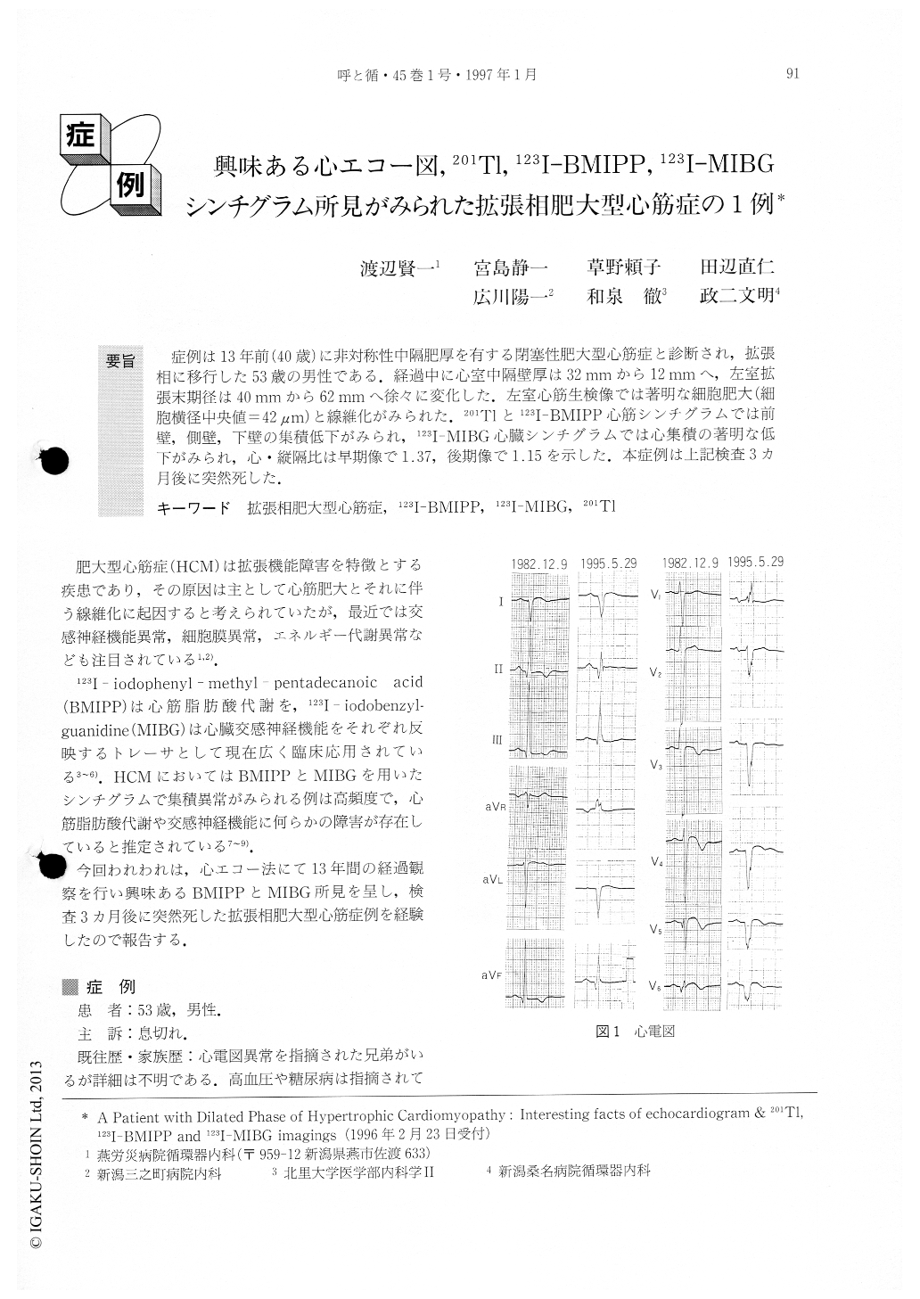
症例は13年前(40歳)に非対称性中隔肥厚を有する閉塞性肥大型心筋症と診断され,拡張相に移行した53歳の男性である.経過中に心室中隔壁厚は32mmから12mmへ,左室拡張末期径は40mmから62mmへ徐々に変化した.左室心筋生検像では著明な細胞肥大(細胞横径中央値=42μm)と線維化がみられた.201Tlと123I-BMIPP心筋シンチグラムでは前壁,側壁,下壁の集積低下がみられ,123I-MIBG心臓シンチグラムでは心集積の著明な低下がみられ,心・縦隔比は早期像で1.37,後期像で1.15を示した.本症例は上記検査3カ月後に突然死した.
We present a case of a 53-year-old male patient who, at the age of 40, was diagnosed as having hypertrophic obstructive cardiomyopathy with asymmetric septal hypertrophy which had progressed to the dilated phase. We were able to observe his condition over a period of 13 years and obtain some intersting facts. The thickness of the intraventricular septum was reduced gradually (from 32 mm to 12 mm) in contrast to the increase in the left ventricular dimension (from 40 mm to 62 mm). The microscopic findings of left ventricular specimens showed marked myocardial hypertrophy (cell diame-ter=42μm) and fibrosis. 201Tl and 123I-iodophenyl-meth-y1-pentadecanoic acid (123I-BMIPP) myocardial images showed marked decreased uptake in anterior, lateral and inferior walls. 123I-iodobenzylguanidine (123I- MIBG) image showed marked decreased heart-to-mediastinum activity ratio (early image=1.37, delayed image=1.15). He died suddenly 3 months after his last hospitalization.

Copyright © 1997, Igaku-Shoin Ltd. All rights reserved.


