Japanese
English
- 有料閲覧
- Abstract 文献概要
- 1ページ目 Look Inside
動脈壁の石灰化は,動脈硬化症を原因とするものがほとんどで,他に血管炎によるものやカルシウム代謝異常によるものなどが知られている。われわれは最近,動脈硬化症のリスク・ファクターを持たず,カルシウム代謝にも異常のみられない若年者に,動脈壁の著明な石灰化と閉塞を伴い,かつほとんど症状のなかったまれな症例を経験したので報告する。
A 14-year-old boy was admitted to our hospital because of abnormal calcification on the X-ray film of his lower extremities. As he had had a transient pain on knee joints after running 5 kilometers, he was taken a X-ray film of knee joints. This revealed bilateral abnormal calcifications along femoral ar-teries from the level just below hip joints to just above knee joints (Fig. 2).
He had never suffered from other diseases except for atopic dermatitis. He was a relatively thin boy (168 cm, 46 kg) with normal features, and no abnormal finding was proved by physical examinations. Nei-ther coldness of extremities nor bruits of vessels was observed. Blood pressure was normal. Although art-eries on upper extremities and at inguinal regions were well palpable, palpation of popliteal and dor-salis pedis arteries was impossible.
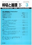
Electrocardiogram and chest X-ray film (Fig. 1) showed no abnormal finding. Laboratory data were also normal except for increased eosinophils and IgE, which could be explained with his atopic dermatitis. We also examined serum levels of calcium, inorganic phosphorus, parathyroid hormones and vitamin D, and all of these data showed normal value.
Angiography was performed and revealed moth-eaten appearances on the external iliac arteries (Fig. 3, arrow) which coincided with the calcifications seen on the plain X-ray film. Bilateral femoral ar-teries were occluded at the level just below hip joints (fig. 3). However, collateral arteries developed well, and gather again at the level of knee joints to form popliteal arteries (Fig. 4).
Other systemic arteries were closely examined, and minute calcified lesions were detected on brachial arteries at elbow joints.
The pattern of calcification observed in this case is different from that of any other calcifying diseases. He is a young boy, and no risk factor for athero-sclerosis could be pointed out from his laboratory data. The X-ray films of his parents were also ex-amined, and no calcification was proved.
As far as we know, no case report like this was published before. We also have to consider the pos-sibility that what we observed in this patient was a process of some established disease (such as pseu-doxanthoma elasticum). So, it is important to follow up him carefully. To find patients similar to this case is also necessary.
Copyright © 1988, Igaku-Shoin Ltd. All rights reserved.