Japanese
English
- 有料閲覧
- Abstract 文献概要
- 1ページ目 Look Inside
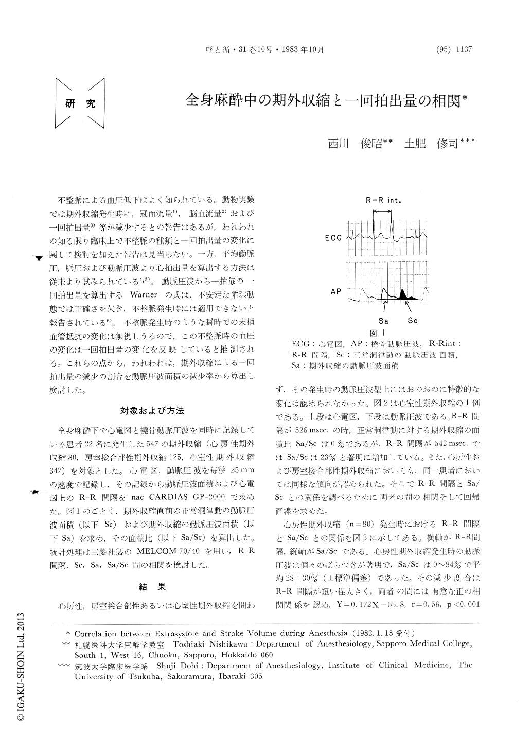
不整脈による血圧低下はよく知られている。動物実験では期外収縮発生時に,冠血流量1),脳血流量2)および一回拍出量3)等が減少するとの報告はあるが,われわれの知る限り臨床上で不整脈の種類と一回拍出量の変化に関して検討を加えた報告は見当らない。一方,平均動脈圧,脈圧および動脈圧波より心拍出量を算出する方法は従来より試みられている4,5)。動脈圧波から一拍毎の一回拍出量を算出するWarnerの式は,不安定な循環動態では正確さを欠き,不整脈発生時には適用できないと報告されている6)。不整脈発生時のような瞬時での末梢血管抵抗の変化は無視しうるので,この不整脈時の血圧の変化は一回拍出量の変化を反映していると推測される。これらの点から,われわれは,期外収縮による一回拍出量の減少の割合を動脈圧波面積の減少率から算出し検討した。
It is speculated that a reduced stroke volume is reflected in a decreased arterial pressure because the peripheral vascular resistance does not change in a moment such as an arrhythmia. The degree of reduced stroke volume with an extrasystole is estimated from the degree of decreased area under the arterial pressure curve above the end-diastolic pressure.
The relationship between Sa/Sc and R-R interval was evaluated, where Sa is an area under the pressure curve above the end-diastolic pressure of an extrasystole, Sc is that of a normal sinus rhythm just before an extrasystole, Sa/Sc is a ratio of Sa and Sc, R-R interval is a time between R wave of a normal sinus rhythm and that of an extrasystole. The shorter the R-R interval was, the greater the degree of the decreased Sa/Sc was. The regression lines obtained in premature atrial and ventricular contractions were Y=0.172 X-55.8 (r=0.56, n=80) and Y=0.217X-72.5 (r=0.75, n=342), respectively.
The above results suggest that a reduced stroke volume with an arrhythmia does not depend on the type of an arrhythmia but mainly on the degree of a decreased left ventricular filling time ; this supports the previous works in animal experiments.

Copyright © 1983, Igaku-Shoin Ltd. All rights reserved.


