Japanese
English
- 有料閲覧
- Abstract 文献概要
- 1ページ目 Look Inside
- 参考文献 Reference
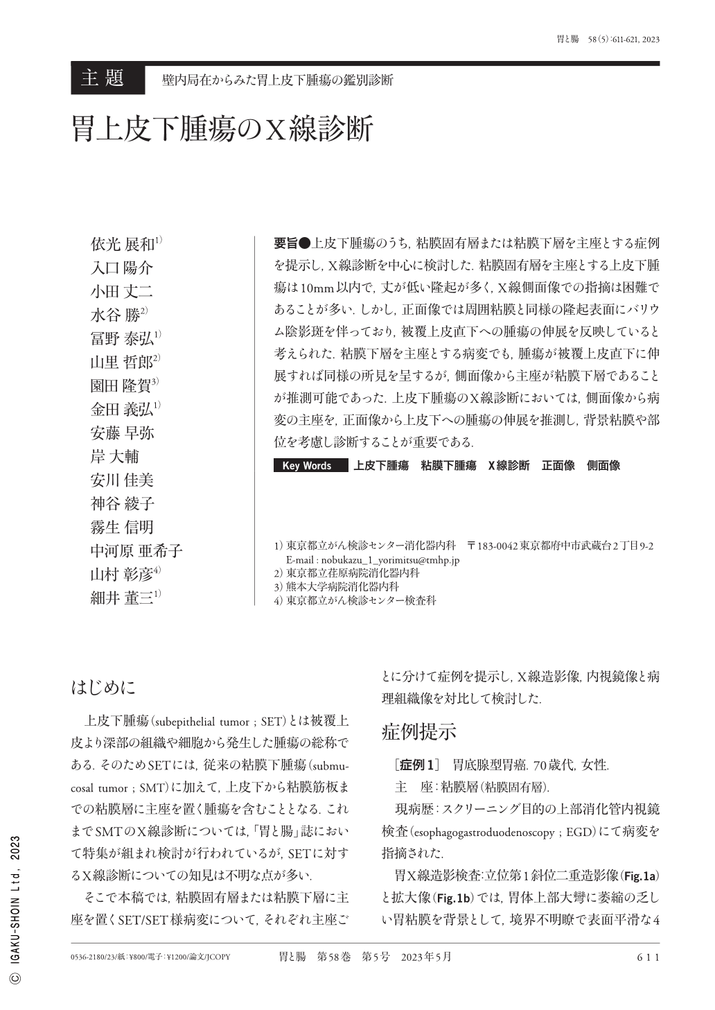
要旨●上皮下腫瘍のうち,粘膜固有層または粘膜下層を主座とする症例を提示し,X線診断を中心に検討した.粘膜固有層を主座とする上皮下腫瘍は10mm以内で,丈が低い隆起が多く,X線側面像での指摘は困難であることが多い.しかし,正面像では周囲粘膜と同様の隆起表面にバリウム陰影斑を伴っており,被覆上皮直下への腫瘍の伸展を反映していると考えられた.粘膜下層を主座とする病変でも,腫瘍が被覆上皮直下に伸展すれば同様の所見を呈するが,側面像から主座が粘膜下層であることが推測可能であった.上皮下腫瘍のX線診断においては,側面像から病変の主座を,正面像から上皮下への腫瘍の伸展を推測し,背景粘膜や部位を考慮し診断することが重要である.
SET(subepithelial tumor)cases emerging from the LPM(lamina propria mucosae)or submucosal layer were presented and reviewed with an emphasis on radiographic diagnosis. The SET-arising from LPM was mainly lower than 10mm, and it is challenging to detect the lesion on the lateral view of upper gastrointestinal contrast imaging. Nevertheless, the frontal view indicated a raised surface similar to the surrounding mucosa with barium shadow spots, reflecting the extension of the tumor just below the covering epithelium. A lesion arising from a submucosal layer would have a similar presentation if the tumor extended just below the covering epithelium. It could thereby be inferred from the lateral view that the SET was in the submucosal layer. In the radiographic diagnosis of SET, it is critical to infer the primary site of the lesion from the lateral view and the extension of the tumor into the subepithelium from the frontal view, considering the background mucosa and site.

Copyright © 2023, Igaku-Shoin Ltd. All rights reserved.


