Japanese
English
- 有料閲覧
- Abstract 文献概要
- 1ページ目 Look Inside
- 参考文献 Reference
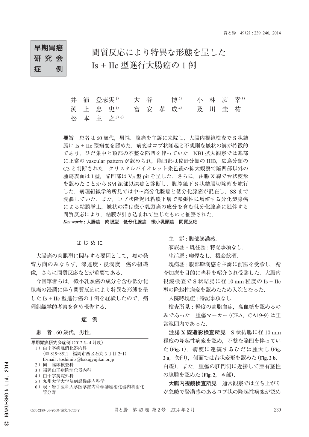
要旨 患者は60歳代,男性.腹痛を主訴に来院し,大腸内視鏡検査でS状結腸にIs+IIc型病変を認めた.病変はコブ状隆起と不規則な皺状の溝が特徴的であり,ひだ集中と頂部の不整な陥凹を伴っていた.NBI拡大観察では基部に正常のvascular patternが認められ,陥凹部は佐野分類のIIIB,広島分類のC3と判断された.クリスタルバイオレット染色後の拡大観察で陥凹部以外の腫瘍表面はI型,陥凹部はVN型pitを呈した.さらに,注腸X線で台状変形を認めたことからSM深部以深癌と診断し,腹腔鏡下S状結腸切除術を施行した.病理組織学的所見では中~高分化腺癌と低分化腺癌が混在し,SSまで浸潤していた.また,コブ状隆起は粘膜下層で膨張性に増殖する分化型腺癌による粘膜挙上,皺状の溝は微小乳頭癌の成分を含む低分化腺癌に随伴する間質反応により,粘膜が引き込まれて生じたものと推察された.
A man in his sixties underwent total colonoscopy for the diagnostic purpose of abdominal discomfort. Conventional colonoscopy revealed a Is+IIc type tumor in the sigmoid colon. Magnifying colonoscopy with the NBI(narrow band imaging)system showed irregular vessels and a non vascular area in the depressed lesion at the top of the tumor . Magnifying colonoscopy with crystal violet showed VN type pit pattern in the depressed lesion at the top of the tumor. Barium enema revealed obvious deformation in the lateral view. Based on these findings, this tumor was diagnosed as the deeply submucosal or more invasive carcinoma. We operated using laparoscopy-assisted sigmoidectomy. Histopathological examination revealed moderately differentiated adenocarcinoma with invasive micropapillary pattern, tub2(>por1>tub1), type 0-Is+IIc, 15×15×4mm, pSS, pN0(0/4), pStage II, med, INFb, ly1(D2-40), ly(+)(SM), v1(VB-HE), v(+)(SM), pPM0(65mm), pDM0(32mm). The interesting shape of this tumor was molded by the desmoplastic reaction.

Copyright © 2014, Igaku-Shoin Ltd. All rights reserved.


