Japanese
English
- 有料閲覧
- Abstract 文献概要
- 1ページ目 Look Inside
- サイト内被引用 Cited by
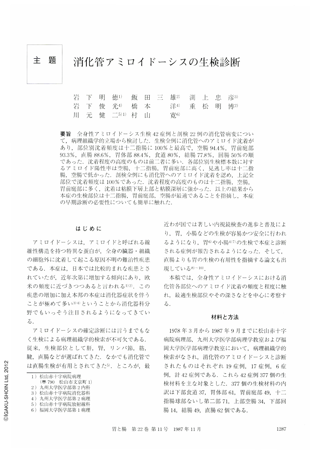
要旨 全身性アミロイドーシス生検42症例と剖検22例の消化管病変について,病理組織学的立場から検討した.生検全例に消化管へのアミロイド沈着があり,部位別沈着頻度は十二指腸に100%と最高で,空腸94.4%,胃前庭部93.3%,直腸88.6%,胃体部88.4%,食道80%,結腸77.8%,回腸50%の順であった.沈着程度の高度のものは前二者に多い.各部位別生検標本数に対するアミロイド陽性率は空腸,十二指腸,胃前庭部に高く,見逃し率は十二指腸,空腸で低かった.剖検全例にも消化管へのアミロイド沈着を認め,上記全部位で沈着頻度は100%であった.沈着程度の高度のものは十二指腸,空腸,胃前庭部に多く,沈着は粘膜下層上部と粘膜深層に強かった.以上の結果から本症の生検部位は十二指腸,胃前庭部,空腸が最適であることを指摘し,本症の早期診断の必要性についても簡単に触れた.
Forty-two biopsy-diagnosed and 22 autopsy cases with systemic amyloidosis were pathologically reviewed with respect to gastrointestinal involvement.
(1) Characteristics of 42 biopsy-diagnosed cases were as folows; average age: 51 years, male to female ratio; 8 to 13, primary amyloidosis: 7 cases, association with multiple myeloma: 4 cases, secondary amyloidosis: 31 cases. On the other hand, characteristics of 22 autopsy cases were as follows; average age: 59.7 years, male to female ratio: 1 to 1, primary amyloidosis: 7 cases, association with multiple myeloma: 5 cases, secondary amyloidosis: 10 cases.
(2) Principal amyloid proteins were AA in 33 cases and AL in 9 cases for biopsy-diagnosis group. Those for autopsy group were AA in 14 cases and AL in 8 cases. Six cases out of 14 primary amyloidosis had AA protein as a principal amyloid constituent.
(3) Gastrointestinal amyloid deposition was observed in all 42 biopsy-diagnosed cases. Sites of amyloid deposition were as follows; duodenum in 100% of cases, jejunum in 94.4%, antrum of the stomach in 93.3%, rectum in 88.6%, body of the stomach in 88.4%, esophagus in 80%, colon in 77.8%, and, least frequently, ileum in 50%. The heaviest deposition was seen in the duodenum and jejum.
(4) Biopsy specimens were divided into two groups depending on whether or not submucosa was included. Amyloid deposition was more frequently observed in the group of biopsy specimens with submucosa taken from most of the gastrointestinal sites. However, submucosa was the only place of amyloid deposition in only 3 cases in that group. Most of the specimens negative for amyloid deposition did not include deep portion of the mucosa.
(5) The proportion of biopsy specimens positive for amyloid among the specimens taken from each site was 94.1% for jejunum, 90.1% for duodenum, 85.7% for antrum of the stomach, 80.6% for rectum, 77% for body of the stomach, 76.9% for esophagus, 47.9% for colon, and 42.9% for ileum.
(6) The proportion of biopsy specimens which were erroneously considered not to have amyloid deposition was 66.7% for ileum, 29.8% for body of the stomach, 21.4% for antrum of the stomach, 20% for esophagus and rectum, 17.4% for colon, 15.6% for duodenum, and 6.3% for jejunum.
(7) Erosive change of the biopsy specimen had no influence on the probability of amyloid deposition. It was detected in 75.5% of the specimens with and in 78.4% of those without erosive change.
(8) Amyloid deposition was noted in the gastrointestinal tract in all 22 autopsy cases. It was true for every site of the tract, i.e., esophagus, stomach, duodenum, jejunum, ileum, colon, and rectum. Duodenum, jejunum, and antrum of the stomach were heavily deposited with amyloid in most of the cases.
(9) In autopsy cases, the chief sites of amyloid deposition in the gastrointestinal tract were, in order of severity, blood vessel walls in the superficial portion of the submucosa and vascular walls with perivascular area in the deep portion of the mucosa.
In conclusion, to diagnose gastrointestinal amyloidosis by biopsy, ①duodenum, antrum of the stomach, and jejunum are best suited, ②mucosa including muscularis mucosa, most ideally submucosa, should be obtained as an appropriate specimen. Brief comment was made on the necessity of early diagnosis of gastointestinal amyloidosis.

Copyright © 1987, Igaku-Shoin Ltd. All rights reserved.


