Japanese
English
- 有料閲覧
- Abstract 文献概要
- 1ページ目 Look Inside
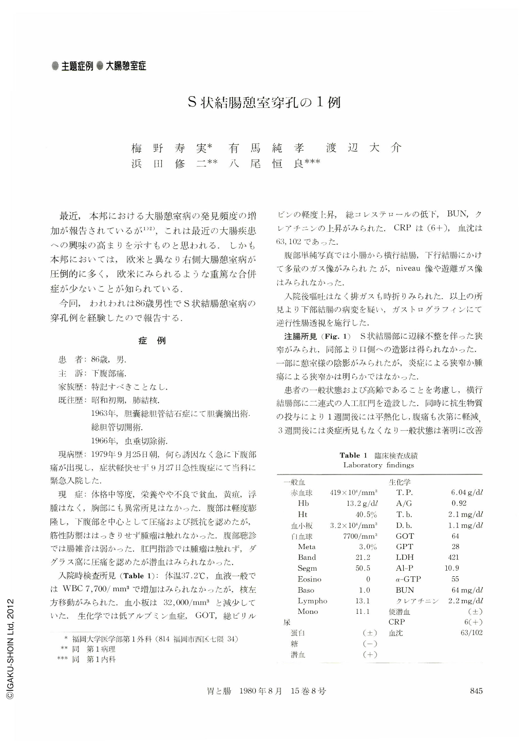
最近,本邦における大腸憩室病の発見頻度の増加が報告されているが1)2),これは最近の大腸疾患への興味の高まりを示すものと思われる.しかも本邦においては,欧米と異なり右側大腸憩室病が圧倒的に多く,欧米にみられるような重篤な合併症が少ないことが知られている.
今回,われわれは86歳男性でS状結腸憩室病の穿孔例を経験したので報告する.
A 86 year-old man entered the hospital complaining of severe lower abdominal pain for two days.
Gastrografin enema on admission revealed the stenosis at the sigmoid colon. Transverse colostomy was performed because of poor condition of the patient. Barium enema a month later showed several diverticula, haustral abnormality and marginal irregularity at the sigmoid colon. The etiology of the stenosis was speculated to be due to acute diverticulitis.
Reoperation was performed at six weeks after colostomy. Purulent coat was observed in the abdominal cavity but the perforated site of gastrointestinal tract was not ascertained. The little finger tip sized several indurations like “sucker of an octopus” were palpated in the wall of the sigmoid colon. Sigmoidectomy was performed and end to end anastomosis was carried out, but transverse colostomy was not closed in order to decrease the pressure affecting the anastomosis site.
Three diverticula, two in the anal side and one in the center, were observed at the resected specimen. Inflammatory redness was observed on the marginal mucosa and the serosal side of the diverticulum in the center and a pin-hole-sized depression was also observed at the reddish serosa.
Histological section of the two diverticula at the anal side showed mucosal herniation with tortuous and thickened muscle layer, measuring 2.0 to 3.0 mm in thickness.
Histologically, findings of the diverticulum in the center were a perforation filled with the granulation tissue with infiltration of inflammatory cells and uniform thickened muscle, 2.1 to 2.3 mm in thickness. On the longitudinal cross section of the resected specimen, muscle abnormality which was an infolding of thickened muscle layer developed toward anal side was seen.

Copyright © 1980, Igaku-Shoin Ltd. All rights reserved.


