Japanese
English
- 有料閲覧
- Abstract 文献概要
- 1ページ目 Look Inside
- サイト内被引用 Cited by
Linitis plastica型の癌は,比較的若い年代に多く,予後も著しく悪い1)10).その多くは胃底腺粘膜領域に小さな原発巣を持つ未分化型癌である3)~5)10).早期胃癌の中に,それを放置しておけばlinitis plastica型に発育する癌が存在することは明らかであるがlinitis plastica型になる癌の初期像の形態は不明である.
癌発生から,あるいは粘膜内癌が粘膜下組織へ浸潤してから,leather bottle状態あるいは胃全体の収縮を示すlinitis plastica型として発見されるまでは,相当長い期間を要していると考えられている2)~4).一方,最近X線学的なlinitis plastica型癌の逆追跡の研究でこの間の形態的変化が明らかにされつつある11)~13).
It has been pointed out that gastric carcinoma of linitis plastics type arises from the fundic gland mucosa without intestinal metaplasia and infiltrates the submucosa antecedently to ulceration in primary focus where cancer cells spread the mucosa. The purpose of this study is to analyse histologically on primary focus of carcinoma having arisen from the fundic gland mucosa.
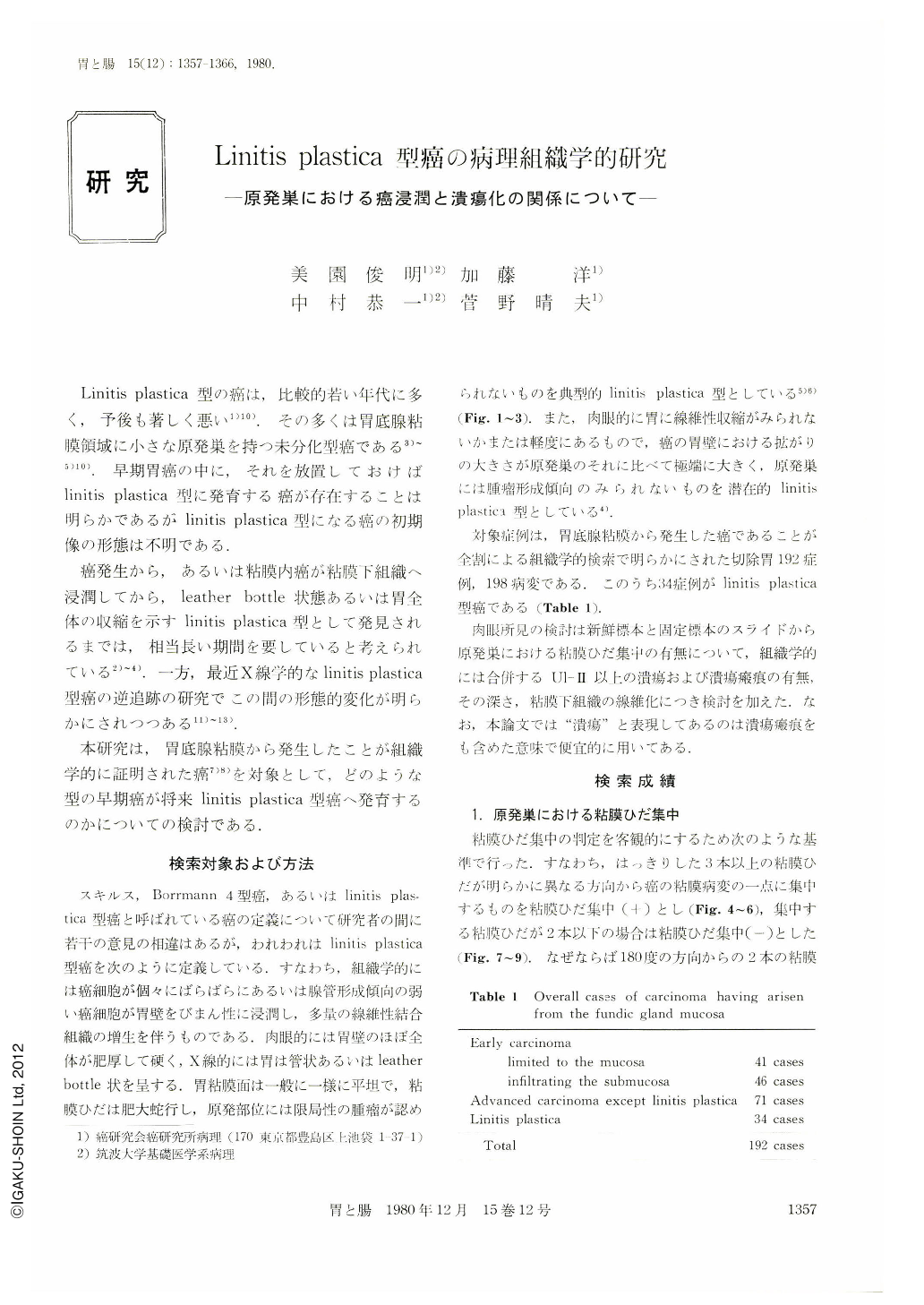
Objects for this study are 192 cases (198 lesions) of gastric carcinoma having arisen from the fundic gland mucosa without intestinal metaplasia. All these resected stomachs were histologically examined by the serially cutting method as shown in Fig. 1~3. Out of the 192 carcinomas, 34 cases are linitis plastics type (Table. 1)
1) Convergency of the mucosal fold in the primary focus : Out of the objects, 151 carcinomas measuring less than 4.0 cm in the largest diameter of the primary focus were examined on convergency of the mucosal fold (Table 2 a). The frequency of the mucosal convergency of the primary focus was less in the linitis plastics type than in other types of gastric carcinoma. Application of Chi-square test between the macroscopic type and convergency of the mucosal fold yields a significant difference as shown in Table 2 b.
Table 4 shows the relationship between convergency of the mucosal fold and ulceration in the primary focus measuring less than 4 cm in diameter. In carcinomas excluding the linitis plastica type, the mucosal convergency is tightly related to the presence of ulceration. It is clear that the mucosal convergency is attributable to contruction of fibrous tissue of ulcerative lesion. Meanwhile, in 23 carcinomas of linitis plastica type measuring less than 4 cm in diameter of the primary focus. 20 primary foci were accompanied with ulcerative lesion. However, 14 out of the 20 linitis plastica type (70%) did not show the mucosal convergency. This finding suggests that cancer cells infiltrated the submucosa antecedently to cancerous ulceration of the primary focus. Because, it is interpreted that the following ulceration is not attributable to convergency of the mucosal fold in area where the muscularis mucosae and the proper muscle were fixed with the intervening fibrosis produced by cancer infiltration.
2) The relationship between depth of co-existing ulcer and size of the primary focus :
Table 5 shows the relationship between depth of ulceration and size of the primary focus. In early and advanced cancer groups except the linitis plastica type, the more the primary focus increases, the deeper the depth of ulcer becomes. These data may show that depth of ulcer co-existing carcinomas becomes gradually deep with the lapse of time. While, the most of the associated ulcerations in linitis plastica type are not deep, independently of the size of the primary focus, as shown in Table 5. It is highly suggested from these data obtained that cancer cells infiltrate the submucosa before the cancerous ulceration in the growing process of carcinoma of the linitis plastica type.
3) The size of the primary focus of the linitis plastica type :
The primary focus is generally small in the linitis plastica type as shown in Table 6. The primary focus measuring less than 2.0 cm occupies 38% in the linitis plastica type, and does 19% in the other advanced cancer. This finding shows that cancer growing to linitis plastica type infiltrated the submucosa in its early phase.
Actually, the undifferentiated carcinoma (diffuse carcinoma) located in the fundic gland mucosa has a striking tendency to infiltrate the submucosa in early phase. Concerning this tendency, Watanabe et al. reported that the undifferentiated carcinoma with submucosal invasion amounted to as much as 70% in this area regardless the largest diameter of the intramucosal spread being under 2.0 cm.
Now, these examinations are large enough to provide a conclusion that the carcinoma growing to linitis plastica type infiltrates the submucosa in its early phase and spreads freely before the cancerous ulceration occurs in the primary focus. Five cases of linitis plastica type which have no ulcerations in the primary focus demonstrate this conclusion (Table 5). The ulcerative fibrosis may play an important role in inhibiting the cancer cells to spread easily in the gastric wall.
According to these consideration, the small carcinoma of Type Ⅱc without convergency of the mucosal fold will grow up to be a linitis plastica type, which carcinoma will be located in the fundic gland mucosa.
Copyright © 1980, Igaku-Shoin Ltd. All rights reserved.