Japanese
English
- 有料閲覧
- Abstract 文献概要
- 1ページ目 Look Inside
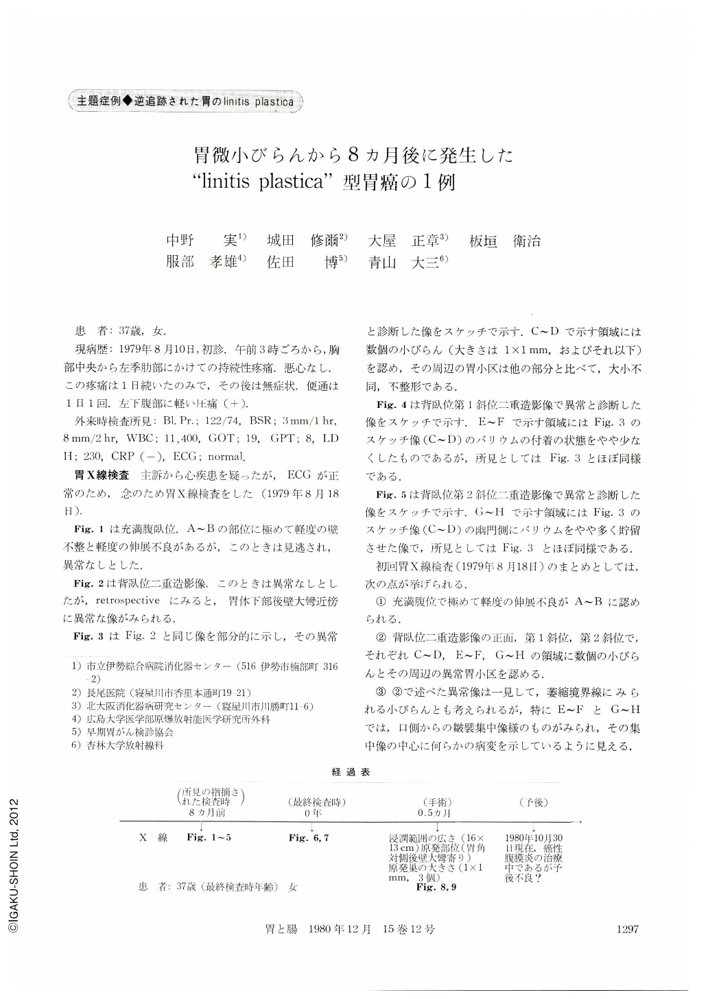
患 者:37歳,女.
現病歴:1979年8月10日,初診.午前3時ごろから,胸部中央から左季肋部にかけての持続性とう痛.悪心なし.この疹痛は1日続いたのみで,その後は無症状.便通は1日1回.左下腹部に軽い圧痛(+).
外来時検査所見:Bl. Pr.;122/74,BSR;3mm/1hr,8mm/2hr,WBC;11,400,GOT;19,GPT;8,LDH;230,CRP(-),ECG;normal.
A 37 year-old woman complained of continuous pain in the middle chest and left hypochondrium since 3 AM, August 10, 1979, and this pain continued during one whole day. After then she had no complaint. Bowel movement once a day. On palpation very slight pain on the left lower abdomen on pressure. Laboratory tests: WBC: 11,400; otherwise within normal limits. ECG : normal.
On August 18, 1979, the first x-ray examination of the stomach was performed. As shown in Figs. 2, 3, 4, and 5, several minute erosions ahout 1 mm in diameter with the surrounding abnormal heteromorph irregular-sized areae gastricae were demonstrated on the posterior wall of the lower body near the greater curvature in the double contrast radiographs in the supine, left posterior and right posterior oblique positions. Those findings may be overlooked at first glance on the routine examinations of many patients, because they are so minute and neither so prominent nor so depressive. Because of those radiological findings, she was recommended to undergo gastrofibroscopy and biopsy in order to confirm histological diagnosis of changes in the gastric mucosa. However, she refused to do so. At that time she had no complaint at all. She did not visit our clinic till April 15, 1980.
Since February 1980, she complained of fullness in the epigastrium during food intake, anorexia, slightly loose stool and slight weight loss, but she had neither nausea nor abdominal pain.
On April 17, 1980, x-ray examination of the stomach was performed as shown in Fig. 6 and 7, depicting the typical form of the linitis plastica type of gastric cancer radiographically.
On April 29, 1980, she was operated upon and the resected specimen of the stomach is shown in Fig. 8. Histologically the typical type of linitis plastica of gastric cancer was observed in Fig. 9.
In the retrospective study of the radiological findings of the initial x-ray examination of the stomach taken on August 18, 1979, the next factors must be carefully remembered.
1) Several minute gastric erosions about 1 mm or less in diameter with the surrounding abnormal heteromorph irregular-sized areae gastricae must be examined with care gastrofibroscopically with biopsy in order to confirm the histological diagnosis of changes in the gastric mucosa.
2) Gastrofibroscopically the diagnosis of minute gastric erosions is not so easy in general as to rule out minute gastric cancer 1 mm or less in diameter.
3) In the stage of minute gastric cancer, rigidity, shadow defect and insufficient distensibility of the gastric wall in the radiological findings are not recognized in general. Even if the above-described radiological findings could not be recognized, gastric cancer must not be ruled out completely.
4) Radiological and gastroscopical diagnosis of minute gastric erosion, small shallow gastric ulcer and abnormal areae gastricae, is very important for the early diagnosis of gastric cancer especially the linitis plastica type of gastric cancer.
5) Clinically doctors observe many x-ray films of the upper gastrointestinal series in general, but minute gastric erosion, small shallow ulcer and abnormal areae gastricae must be more carefully diagnosed to rule out the early stage of gastric cancer.
Finally the gastroscopical diagnosis for minute gastric erosion, if the radiological abnormalities were recognized, must be more carefully performed. When the negative results were reported in biopsy, repeated precise examination of x-ray and gastroscopy must be performed every month and the patient must be followed up very carefully.
Copyright © 1980, Igaku-Shoin Ltd. All rights reserved.